


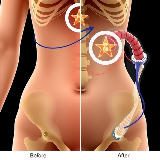
A second weight-loss operation may carry an overlooked cost for the skeleton, according to a new study of postmenopausal women. Researchers in France found that women who underwent a revisional Roux-en-Y gastric bypass after first having sleeve gastrectomy had substantially lower bone mineral density at the hip and femoral neck than women who underwent Roux-en-Y bypass as their initial bariatric procedure. The difference persisted even after the investigators accounted for age, body-mass index and the time elapsed since surgery. The findings, published in Archives of Osteoporosis, suggest that the type and sequence of bariatric operations may matter for long-term skeletal health, not only for weight loss and metabolic improvement. For patients and clinicians, the result raises an urgent question: could a procedure performed to improve the outcome of an earlier operation also intensify vulnerability to osteoporosis and fractures later in life?
Bariatric surgery is among the most effective treatments for severe obesity, reducing body weight and often improving type 2 diabetes, high blood pressure, sleep apnoea and other obesity-related conditions. But the operations can alter the body’s handling of nutrients, hormones and mechanical forces in ways that affect bone. Sleeve gastrectomy removes much of the stomach, creating a narrow tube and reducing food intake, while Roux-en-Y gastric bypass both restricts intake and reroutes food around the stomach and the first part of the small intestine. That rerouting can reduce the absorption of calcium and vitamin D, nutrients essential for maintaining mineralised bone. Rapid weight loss also decreases the loading forces transmitted through the skeleton, potentially reducing the stimulus that normally helps preserve bone mass. Changes in gut hormones and bone-remodelling signals may add to the effect.
The new study focused on 120 postmenopausal women treated at a single centre: 97 had undergone primary Roux-en-Y gastric bypass, and 23 had received the operation as a revision after sleeve gastrectomy. Revisional surgery is performed for several reasons, including inadequate weight loss, weight regain, reflux or other complications after the original operation. It is not simply a repeat of the first procedure. The patient has already experienced one period of weight loss and anatomical alteration, and the second operation may produce an additional shift in nutrient absorption and gastrointestinal physiology. Postmenopausal women are a particularly important group for studying bone because declining oestrogen levels accelerate bone turnover and increase the risk of osteoporosis. The researchers compared demographic characteristics, medical conditions and recognised osteoporosis risk factors before examining the women’s skeletal and body-composition measurements.
Bone mineral density was measured using dual-energy X-ray absorptiometry, or DXA, the standard clinical technique for estimating bone mass. DXA uses two low-dose X-ray beams to distinguish bone mineral from soft tissue, producing measurements in grams per square centimetre. It also generates T-scores, which compare a patient’s bone density with the average density of a healthy young adult. A T-score between −1.0 and −2.5 is generally classified as low bone mass, or osteopenia, while a score of −2.5 or below is consistent with osteoporosis, although diagnosis also depends on clinical context and fracture history. In this study, scans assessed the lumbar spine, femoral neck and total hip. The same examinations also estimated fat mass and lean mass, allowing the investigators to determine whether differences in bone density simply reflected differences in body size or muscle and fat composition.
Before statistical adjustment, the revisional-surgery group had a mean femoral-neck bone mineral density of 0.693 grams per square centimetre, compared with 0.761 grams per square centimetre among women who had primary bypass surgery. This difference was statistically significant, with a P value of 0.011. The corresponding average T-scores were −1.4 and −0.9, respectively, a difference that was also significant. In practical terms, the revisional group’s average femoral-neck measurement fell further into the low-bone-mass range, although the study does not report that every individual patient had osteopenia or osteoporosis. Initial comparisons found no meaningful differences in body-mass index, major comorbidities or recorded osteoporosis risk factors. Bone density at the lumbar spine and total hip appeared similar before adjustment, showing that the signal was most pronounced at the femoral neck.
The more revealing result emerged when the team adjusted the analysis for age, body-mass index and time since surgery. After this correction, total-hip bone mineral density remained lower in the revisional group, with an adjusted mean of 0.834 grams per square centimetre, compared with 0.899 grams per square centimetre in the primary-surgery group. The P value was 0.017, and the 95 per cent confidence intervals were 0.786 to 0.882 and 0.877 to 0.921, respectively. At the femoral neck, adjusted density averaged 0.682 grams per square centimetre after revision and 0.763 grams per square centimetre after primary bypass, with a P value of 0.003. Because the confidence intervals and statistical tests indicate a persistent group difference, the findings are unlikely to be explained solely by the women being older, having different body sizes or simply having undergone surgery at different times.
The absence of a body-composition difference is scientifically important. The researchers found similar proportions of fat and lean tissue in the two groups, so the lower hip density after revisional surgery could not be readily attributed to a greater loss of muscle or a markedly different physical build. Nevertheless, the study cannot establish that revisional surgery directly caused the bone loss. Its cross-sectional design means that participants were assessed after their operations rather than followed from before surgery through successive years. The women may have differed in unmeasured ways, such as the amount of weight they lost after each procedure, dietary intake, calcium and vitamin-D supplementation, physical activity, smoking, alcohol use, medication exposure or pre-existing bone density. The small revisional group, comprising only 23 women, also limits the precision with which the results can be generalised.
The biological explanation is plausible, however. Bone is living tissue that is continuously remodelled: osteoclasts remove old or damaged bone, while osteoblasts form new tissue. When resorption outpaces formation, mineral density declines. After Roux-en-Y bypass, reduced exposure of nutrients to the duodenum and proximal jejunum can impair calcium absorption. A fall in absorbed calcium may stimulate parathyroid hormone, which helps maintain blood calcium partly by increasing calcium release from bone. Vitamin-D deficiency can compound the problem by reducing intestinal calcium uptake. At the same time, weight loss reduces skeletal loading, and altered concentrations of gut hormones such as glucagon-like peptide-1 and peptide YY may influence bone turnover. A revisional bypass could expose patients to overlapping or repeated metabolic stresses, although the present study did not measure all these pathways or prove that any one mechanism accounted for the hip findings.
The location of the difference may also matter. The femoral neck and total hip are weight-bearing regions that respond strongly to changes in body mass, muscle forces and physical activity. The lumbar spine, by contrast, can be affected by degenerative changes that sometimes make DXA readings appear higher than the amount of healthy bone would suggest, particularly in older adults. Lower density at the hip is clinically concerning because hip fractures can lead to prolonged loss of independence, complications and increased mortality in older people. Yet a low DXA value is not itself a fracture forecast, and the study did not report fracture outcomes. It therefore signals a risk that merits attention rather than demonstrating that revisional bypass inevitably leads to fractures. The researchers’ conclusion is more measured: women undergoing revisional procedures may need targeted, long-term skeletal monitoring.
That monitoring could include assessment of fracture history and risk factors, periodic DXA scans when clinically appropriate, and laboratory evaluation of calcium, vitamin D and related markers. Nutritional follow-up is already a central part of bariatric care, but the findings suggest that skeletal surveillance should be especially deliberate after a sleeve-to-bypass conversion. Clinicians may need to review whether patients are taking prescribed supplements, absorbing them adequately and maintaining sufficient protein and calcium intake, while also encouraging safe resistance and weight-bearing exercise tailored to individual ability. Decisions about osteoporosis medication require specialist judgement because altered gastrointestinal anatomy and nutritional status can affect treatment choices. The study does not argue against revisional bariatric surgery, which can provide substantial health benefits, nor does it show that primary bypass is harmless to bone. Instead, it adds a warning to the rapidly expanding medical conversation around obesity treatment: success measured on the bathroom scale may conceal changes in tissues that are harder to see.
Further research will need to follow larger groups of patients before and after both operations, measure bone-turnover markers and nutrient levels, and track fractures over time. Direct comparisons should also examine whether the interval between sleeve gastrectomy and revision, the length of the bypassed intestine, the magnitude of weight loss or the use of anti-obesity medicines changes skeletal outcomes. Randomised trials of supplementation, exercise programmes or bone-protective therapies could reveal how much of the decline is preventable. For now, the French study offers one of the clearest indications that revisional Roux-en-Y gastric bypass may affect postmenopausal bone differently from the same operation performed as a first procedure. As bariatric surgery becomes more common and more patients undergo sequential operations, the hip—not just the waistline—may become an essential part of measuring long-term success.
Cite Scienmag News
Ophelia Keating. (August 27, 2026). Revisional Roux-en-Y bypass after sleeve gastrectomy affects bone health in postmenopausal women. Scienmag. https://scienmag.com/revisional-roux-en-y-bypass-after-sleeve-gastrectomy-affects-bone-health-in-postmenopausal-women/
Ophelia Keating. "Revisional Roux-en-Y bypass after sleeve gastrectomy affects bone health in postmenopausal women." Scienmag, 27 August 2026, https://scienmag.com/revisional-roux-en-y-bypass-after-sleeve-gastrectomy-affects-bone-health-in-postmenopausal-women/. Accessed 5 September 2026.
Ophelia Keating. "Revisional Roux-en-Y bypass after sleeve gastrectomy affects bone health in postmenopausal women." Scienmag. August 27, 2026. https://scienmag.com/revisional-roux-en-y-bypass-after-sleeve-gastrectomy-affects-bone-health-in-postmenopausal-women/


