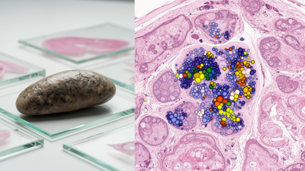
High-resolution mass-spectrometry imaging has produced an organ-level lipid atlas of the common liver fluke, Fasciola hepatica, showing that lipids associated with the parasite’s surface, intestine and reproductive organs occupy distinct locations within adult worms. The study used atmospheric-pressure scanning microprobe matrix-assisted laser desorption/ionization mass spectrometry imaging, or AP-SMALDI MSI, coupled to an Orbitrap analyzer, and complemented the spatial measurements with bulk liquid chromatography tandem mass spectrometry. Among 377 reproducibly detected signals in positive-ion mode and 150 in negative-ion mode, the researchers assigned 73% and 61%, respectively, of previously database-annotated signals to lipid species supported by LC-MS/MS. The experiments involved laboratory-reared parasites obtained from rats, not samples collected from infected people or livestock.
Fasciola hepatica causes fascioliasis, a food-borne neglected tropical disease affecting humans and grazing animals across all inhabited continents. Triclabendazole is the only medically approved drug for human fascioliasis, and more frequent reports of resistance have increased interest in the parasite’s molecular biology and possible intervention targets. Lipids contribute to membrane structure, energy storage, cell signaling and parasite-host interactions, yet much of the earlier information about this parasite came from bulk tissue analyses that could not show where individual compounds occurred.
The new work instead maps molecular signals across four organs central to host contact and reproduction: the tegument, intestine, ovary and testis. For the imaging experiments, researchers analyzed transverse sections measuring 20 micrometres thick as well as the native surface of whole worms. AP-SMALDI MSI used a pixel size of 7 or 8 micrometres, a mass range of m/z 250–1,000 and a mass resolving power of 240,000 at m/z 200.
Pixel-wise autofocus was particularly important for native specimens: one surface measurement lasted four hours, during which moisture loss lowered the sample by about 500 micrometres. Without continual adjustment of the laser focus, the authors report, signal intensity would have declined until signals were lost entirely. Distinct matrix compounds were used for positive- and negative-ion analysis, and selected tissue sections later underwent hematoxylin and eosin staining so that mass-spectral regions could be checked against anatomical structures.
The researchers established reproducible signal lists for each organ and ion mode. A signal had to occupy at least 5% of the pixels in its region of interest, fall within the mass range used to focus on lipids, and exceed an average intensity of 150, while also appearing in at least three of four biological replicates. These signals were searched against the LIPIDMAPS Structure Database and then compared with bulk LC-MS/MS results.
The workflow assigned signals to five levels of identification confidence, ranging from signals without a database match to those with MS/MS evidence that resolved more than bulk structure, including individual fatty-acid composition. Even at the higher levels, the method did not determine double-bond positions or the sn-position of fatty-acyl chains. The approach was therefore designed to produce a reproducible map of lipid distributions and candidate identities, not an exact catalogue of every molecular isomer.
The resulting maps showed clear chemical differences among the four organs. Principal component analysis separated the tegument, intestine, ovary and testis in both ionization modes, with the tegument appearing particularly distinctive in positive-ion mode. The surface was enriched in ceramide-related species, including hexosylceramides and inositol phosphoryl ceramides, and also contained a high proportion of lysophospholipids and ether-linked glycerophospholipids.
The intestine likewise contained many ceramide-related lipids, indicating a shared but non-identical chemical profile for the two epithelial structures that contact the host. In contrast, the gonads were characterized by unsaturated phosphatidylinositol species, whereas saturated phosphatidylinositol signals such as PI 32:0 and PI 34:0 were prominent in the tegument. These are observed spatial distributions; the imaging data do not establish that any particular lipid causes a particular physiological function.
At 7-micrometre resolution, the researchers also distinguished the intestinal lumen from the gastrodermis that surrounds it. The lumen exclusively contained signals assigned to hexosylceramides, inositol phosphoryl ceramides and phosphatidylserines, whereas the gastrodermis included phosphatidylcholines, phosphatidylethanolamines, dimethylphosphatidylethanolamines, phosphatidic acids, phosphatidylinositols and diglycerides with matching carbon-chain lengths. Differences in ceramide hydroxylation were also apparent: Cer 36:1;O2 occurred in the intestine, HexCer 38:0;O3 occurred in both intestine and tegument, and HexCer 38:0;O4 was detected in the tegument.
These findings provide a chemical distinction between anatomical compartments that could not be captured by bulk extraction alone, although the study did not directly establish the function of the compounds in either compartment. Native-surface imaging exposed additional regional variation within the tegument. Across technical and biological replicates, the affected signals were two to nine times more intense ventrally, although principal component analysis did not clearly separate the two sides, indicating a quantitative rather than wholesale compositional difference.
Some signals associated with ether-linked lipids and saturated phosphatidylinositols co-localized in the tegument and its underlying cyton layer. The researchers cautioned that a spine-shaped pattern in positive-ion images likely reflected crystallization of the matrix on the uneven surface rather than a biological structure. The native worm therefore required careful interpretation because changing sample height and uneven matrix deposition could influence apparent spatial patterns.
The reproductive organs showed partly shared and partly distinct lipid profiles. Both testis and ovary contained unsaturated phosphatidylcholines, phosphatidylethanolamines, phosphatidylinositols and corresponding diglycerides, including common species such as PC 40:4 and PC 40:5. The ovary additionally accumulated PC 42:4, while PC 38:3 occurred in the ovary, vitellarium and gastrodermis and was more abundant around the ovary’s outer margin.
The authors proposed that some patterns may correspond to distinct ovarian cell populations, but the imaging data alone did not establish those identities or functions. The maps therefore identify candidate tissue associations for follow-up experiments rather than demonstrate a cell-type-specific molecular mechanism. The survey also identified unusual molecular features.
F. hepatica contained lipids with odd-numbered carbon chains, including PC 31:0, PC 33:0, PE 35:1 and PS 43:4, as well as very-long-chain components reaching at least 22 and 24 carbons in directly annotated species; LC-MS/MS further suggested components as long as 26–32 carbons. Triglycerides such as TG 54:6, TG 56:6 and TG 58:10 shared a broad distribution across parenchymatic tissue, while other glycerophospholipid signals occupied localized structures that might correspond to excretory ducts. The authors noted that the functions of odd-chain and very-long-chain fatty-acid components in this parasite remain unclear.
The imaging and LC-MS/MS datasets, together with organ-specific signal lists and spatial maps, were deposited in public repositories, making the atlas a reference resource for studies of parasite physiology, host interaction and molecular drug research. Several limitations constrain interpretation. The measurements were not quantitative, and the workflow could not determine double-bond positions or sn-position isomers, so many assignments describe lipid class, carbon number and saturation rather than a fully resolved molecular structure.
Because the LC-MS/MS analysis used bulk worm extracts, isobaric lipids from other tissues could have been co-attributed to signals assigned to an organ. Regions of interest were selected with mass-spectral markers rather than perfect anatomical boundaries and may include a small fraction of neighboring tissue, while structures smaller than the 7–8-micrometre lateral resolution were not distinguishable. The study nevertheless demonstrates that label-free, spatially resolved lipidomics can map organ-associated chemical patterns in an adult parasitic worm.
Its principal contribution is a measured tissue atlas that future functional studies can use to test whether particular lipids contribute to barrier function, signaling, reproductive biology or parasite-host interaction; it is not evidence of a new treatment or clinical benefit.
Subject of Research: Spatial lipid profiling of the tegument, intestine, ovary and testis in adult Fasciola hepatica
Article Title: Spatial lipidomics of Fasciola hepatica using high-resolution AP-SMALDI Orbitrap MSI – a lipid tissue atlas
Article References: Morawietz, C. M., Gerbig, S., Haeberlein, S., & Spengler, B. (2026). Spatial lipidomics of Fasciola hepatica using high-resolution AP-SMALDI Orbitrap MSI – a lipid tissue atlas. Frontiers in Cellular and Infection Microbiology, 16, 1934046. https://doi.org/10.3389/fcimb.2026.1934046
Image Credits: AI Generated
DOI: 10.3389/fcimb.2026.1934046
Keywords: Fasciola hepatica, spatial lipidomics, MALDI mass spectrometry imaging, AP-SMALDI MSI, Orbitrap mass spectrometry, LC-MS/MS, parasite lipids, liver fluke, fascioliasis, tissue atlas, Spatial, lipidomics
Cite Scienmag News
Drew Townsend. (October 22, 2026). Research report: Spatial lipidomics of Fasciola hepatica using high-resolution AP-SMALDI Orbitrap MSI – a lipid tissue atlas. Scienmag. https://scienmag.com/research-report-spatial-lipidomics-of-fasciola-hepatica-using-high-resolution-ap-smaldi-orbitrap-msi-a-lipid-tissue-atlas/
Drew Townsend. "Research report: Spatial lipidomics of Fasciola hepatica using high-resolution AP-SMALDI Orbitrap MSI – a lipid tissue atlas." Scienmag, 22 October 2026, https://scienmag.com/research-report-spatial-lipidomics-of-fasciola-hepatica-using-high-resolution-ap-smaldi-orbitrap-msi-a-lipid-tissue-atlas/. Accessed 22 October 2026.
Drew Townsend. "Research report: Spatial lipidomics of Fasciola hepatica using high-resolution AP-SMALDI Orbitrap MSI – a lipid tissue atlas." Scienmag. October 22, 2026. https://scienmag.com/research-report-spatial-lipidomics-of-fasciola-hepatica-using-high-resolution-ap-smaldi-orbitrap-msi-a-lipid-tissue-atlas/