

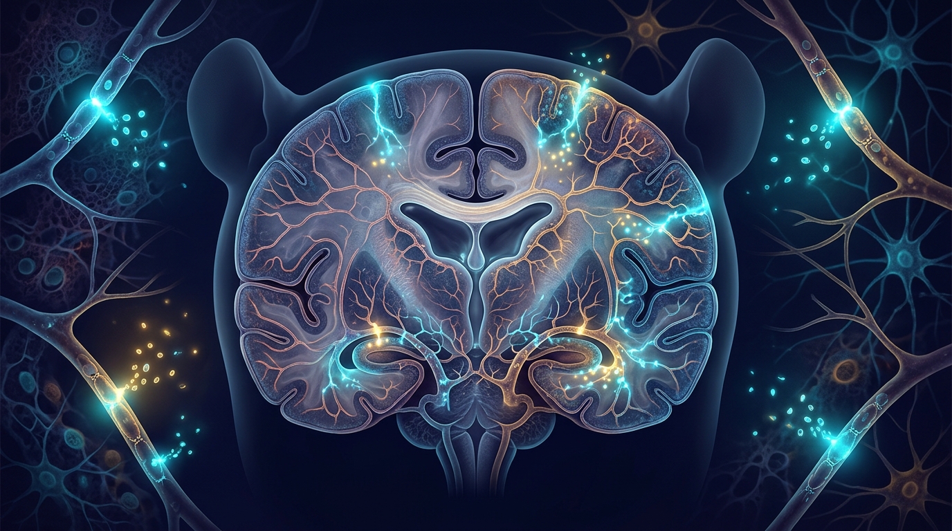
A single genetic change can push a rat’s brain fluid system out of balance, and the barrier that normally keeps blood components out of brain tissue appears to leak across multiple regions at once. Researchers report in Fluids and Barriers of the CNS that rats carrying a truncating mutation in the Cwh43 gene develop enlarged brain ventricles, impaired motor coordination and measurable blood-brain barrier permeability abnormalities spanning cortical, hippocampal and cerebellar tissue. The work links a disease-associated genetic factor to regional barrier phenotypes in an experimental model of idiopathic normal pressure hydrocephalus, an aging-associated neurological disorder characterized by ventricular enlargement and disturbances in brain fluid homeostasis.
The team generated the model using CRISPR/Cas9 genome editing. The mutant allele was defined as Cwh43 NM_001191677.1:c.1597del, which predicts a truncated protein designated NP_001178606.1:p.(Met533Ter). Rats carrying this allele showed reduced Cwh43 transcript abundance. Heterozygous mutant animals developed ventricular enlargement and performed worse on the rotarod test, a standard measure of motor coordination and balance. These features mirror core aspects of normal pressure hydrocephalus, a condition in which the brain’s fluid-filled chambers expand despite measurements of cerebrospinal fluid pressure that fall within expected ranges.
To probe the barrier directly, the researchers injected tracers of three different molecular weights into the bloodstream: small molecules of 556 daltons, mid-sized proteins of 10 kilodaltons and large proteins of 70 kilodaltons. Repeated-measures analyses of the three tracer cohorts demonstrated significant overall genotype-associated blood-brain barrier permeability phenotypes across a multiregional dataset. In practical terms, the amount of tracer that escaped from blood vessels into surrounding brain tissue differed between mutant and control animals in a pattern that held across regions as distinct as the cortex, the hippocampus and the cerebellum.
What makes the finding structurally interesting is what the team did not observe. The permeability phenotypes occurred without a significant reduction in the measured full-length junctional proteins, the molecular components that physically tether neighboring endothelial cells together at the barrier. Representative ultrastructural images obtained by transmission electron microscopy showed identifiable vascular junctional contacts. The barrier therefore does not appear to fail by simply losing its visible junctional hardware, at least not in ways captured by the protein measurements and imaging performed in this cross-sectional study.
The mutant rats also displayed signs of vascular and ventricular-interface stress. Vessel-associated VCAM-1 immunoreactivity, a marker of endothelial activation, was increased in mutant animals. However, whole-tissue VCAM-1 and the measured inflammasome-associated proteins did not show consistent genotype-dependent increases, suggesting the inflammatory signal is localized to the vessel wall rather than spread diffusely through the tissue. Additional analysis identified reduced apical acetylated α-tubulin-positive coverage and mean fluorescence intensity along the lateral ventricular ependymal surface, the cellular lining that faces the cerebrospinal fluid and helps maintain the interface between brain tissue and ventricular contents.
In the periventricular region adjacent to the ventricles, normalized tracer values were higher in heterozygous mutant rats across all three tracer cohorts. Yet none of the individual genotype comparisons in that region remained significant after correction for the three-tracer family, a statistical safeguard that accounts for testing multiple tracers simultaneously. The researchers report the trend but treat it cautiously, distinguishing the multiregional overall phenotype, which survived repeated-measures analysis, from the periventricular pattern, which did not withstand multiple-comparison correction on its own.
The study’s design matters for interpreting these results. This is a cross-sectional experimental model, not a longitudinal study of disease progression. The authors state explicitly that the present cross-sectional study does not establish the temporal ordering of the observed phenotypes, their primary cellular origin, or the underlying molecular mechanism. In other words, the work cannot say whether the barrier leak precedes ventricular enlargement, follows it, or develops in parallel through a shared upstream cause. Nor can it identify which cell type initiates the cascade of vascular, glial and ventricular-interface changes documented in the mutant animals.
What the model does provide is a framework. By tying a specific truncating allele in Cwh43 to reproducible multiregional barrier permeability phenotypes alongside vascular, glial and ventricular-interface abnormalities, the researchers have created an experimental platform for dissecting how genetic risk factors translate into barrier dysfunction in normal pressure hydrocephalus. The combination of tracer-based permeability quantification, immunoblotting, immunofluorescence and transmission electron microscopy offers multiple independent readouts for future mechanistic studies in CWH43-related experimental models.
For a field in which experimental models linking disease-associated genetic factors to regional barrier phenotypes have remained limited, the value of this work lies in its specificity. The mutation is precisely defined at the nucleotide and protein level, the permeability measurements span three tracer sizes and multiple brain regions, and the statistical treatment distinguishes robust genotype-associated phenotypes from trends that do not survive correction. The result is a carefully bounded but reproducible experimental signature of barrier and ventricular-interface disruption in a genetically defined rat model of a disorder that primarily affects older adults.
Subject of Research: Blood-brain barrier permeability abnormalities in a Cwh43 p.(Met533Ter) rat model of normal pressure hydrocephalus
Article Title: Multiregional blood-brain barrier permeability abnormalities in a Cwh43 truncating-allele p.(Met533Ter) rat model of normal pressure hydrocephalus
Article References: Zhu, Q., Yan, Z., Li, M., Wang, J., Wu, X., Cao, J., Wei, Z., Zhang, J., Liu, Y., Chen, Z., Cheng, Y., Zheng, Y., Zhang, H., Huang, M., Chen, H., Cao, H., Hu, C., Li, A., & Yang, D. (2026). Multiregional blood-brain barrier permeability abnormalities in a Cwh43 truncating-allele p.(Met533Ter) rat model of normal pressure hydrocephalus. Fluids and Barriers of the CNS. https://doi.org/10.1186/s12987-026-00900-8
Image Credits: AI Generated
DOI: 10.1186/s12987-026-00900-8
Keywords: normal pressure hydrocephalus, blood-brain barrier, Cwh43, CRISPR/Cas9, rat model, ventricular enlargement, VCAM-1, ependymal surface, tracer permeability, rotarod, transmission electron microscopy, Fluids and Barriers of the CNS
Cite Scienmag News
Cassandra Pierce. (November 4, 2026). Rat model of normal pressure hydrocephalus shows multiregional blood-brain barrier leaks. Scienmag. https://scienmag.com/rat-model-of-normal-pressure-hydrocephalus-shows-multiregional-blood-brain-barrier-leaks/
Cassandra Pierce. "Rat model of normal pressure hydrocephalus shows multiregional blood-brain barrier leaks." Scienmag, 4 November 2026, https://scienmag.com/rat-model-of-normal-pressure-hydrocephalus-shows-multiregional-blood-brain-barrier-leaks/. Accessed 4 November 2026.
Cassandra Pierce. "Rat model of normal pressure hydrocephalus shows multiregional blood-brain barrier leaks." Scienmag. November 4, 2026. https://scienmag.com/rat-model-of-normal-pressure-hydrocephalus-shows-multiregional-blood-brain-barrier-leaks/