


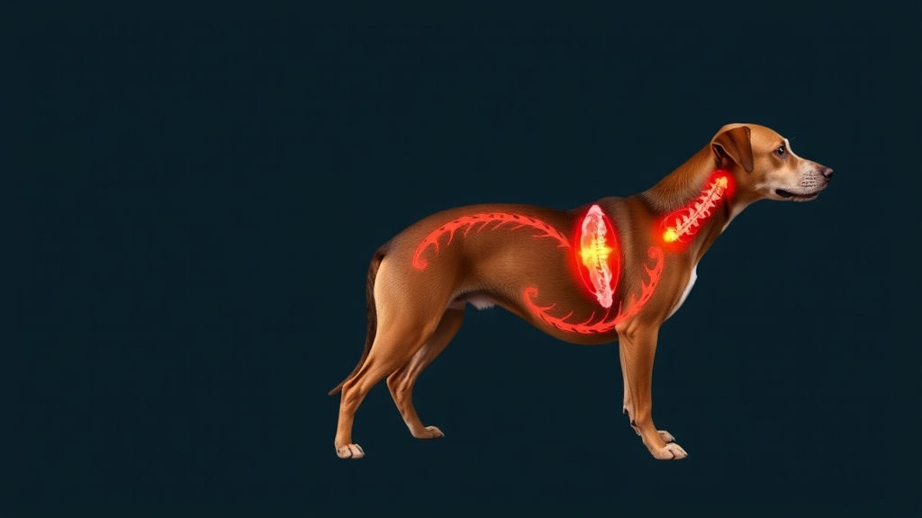
Mast cell tumours are the most familiar villain in canine dermatology, the single most common malignant skin cancer in dogs, accounting for an estimated 11 to 18 percent of all cutaneous neoplasms in the species. Most veterinarians know them as lumps in or under the skin, sometimes solitary, sometimes multiple, occasionally invasive and capable of seeding distant organs. What almost nobody expects to see is a mast cell tumour lighting up the spine in multiple vertebrae at once. A new case series published in the open-access journal Veterinary Oncology documents exactly that rare scenario in three dogs, and its findings could change how veterinary neurologists and oncologists interpret aggressive vertebral lesions on magnetic resonance imaging.
The report, led by Freya Townsend of Wear Referrals Veterinary Hospital in Stockton-on-Tees, United Kingdom, describes three dogs in which metastatic mast cell tumours produced polyostotic vertebral lesions, meaning tumour deposits scattered across multiple vertebrae rather than a single site of bone destruction. According to the authors, only one previous report in the veterinary literature has documented polyostotic mast cell tumour metastasis affecting the vertebrae. That scarcity is precisely what makes the new series noteworthy: it suggests that mast cell tumours deserve a place on the differential diagnosis list for extradural and polyostotic vertebral disease, a consideration that could influence biopsy decisions, staging protocols and prognostic conversations with owners.
The first case involved an eight-year-old neutered male small crossbreed presented originally for a small, raised mass on the upper lip. Histopathology classified it as a grade II tumour under the traditional Patnaik system and low grade under the more prognostically stringent Kiupel system, with a low mitotic count of one mitosis per ten high-power fields. The mass was excised completely, albeit with a narrow deep margin. Seven months later, the right mandibular lymph node was enlarged and biopsy confirmed metastatic mast cell disease. Fine needle aspirates of the opposite lymph node, liver and spleen were reassuringly negative at that stage, and the dog was started on lomustine chemotherapy. When a subcutaneous nodule later appeared at the surgical site and cytology again confirmed metastasis, treatment was switched to toceranib phosphate before referral to a specialist oncology service.
At the referral hospital, computed tomography revealed enlargement of the right mandibular and bilateral medial retropharyngeal lymph nodes, and surgical removal of these nodes confirmed overt metastatic mast cell infiltration, graded HN3 under the Weishaar classification system for nodal mast cell metastasis. The dog then received vinblastine chemotherapy combined with prednisolone, followed by a five-day course of radiotherapy totalling 20 Gy to the surgical site. One week after radiotherapy ended, the dog became reluctant to exercise. Neurological examination revealed pain on palpation of the thoracolumbar region, and within another week the dog had developed a hunched spine and ambulatory paraparesis, with absent postural reactions in the pelvic limbs. Blood work showed elevated C-reactive protein alongside neutropenia, leukopenia and reduced platelets, consistent with both inflammatory disease and chemotherapy side effects.
The second case was a fifteen-year-old neutered female springer spaniel whose story began with removal of a two-centimetre subcutaneous mass from the right caudal mammary gland. That tumour carried a high mitotic count exceeding ten mitoses per ten high-power fields, and the regional lymph node was already invaded by sheets of hyperchromatic, intermediately differentiated mast cells, classified HN2. The mass tested negative for a c-KIT mutation. Seven months after surgery, a new groin mass and imaging findings of enlarged abdominal lymph nodes and liver nodules confirmed widespread metastasis, and vinblastine with prednisolone was initiated. Two months into chemotherapy, the dog developed ambulatory paraparesis that progressed to non-ambulatory paraparesis by the time of specialist assessment, with pain suspected on palpation of the cervical spine and neurological signs localising to the mid-thoracic spinal cord segments.
The third patient, a thirteen-year-old neutered male Nova Scotia Duck Tolling Retriever, had a long history of mast cell tumours, including a recurrent low-grade shoulder mass and, more recently, a twenty-millimetre high-grade Kiupel tumour on the mid back with narrow surgical margins. Abdominal ultrasound had already shown nodular changes in the liver and spleen. The dog completed a twelve-week course of vinblastine and prednisolone but skipped restaging, and seven months later returned with a one-week history of non-ambulatory paraparesis, a large thoracic wall mass in the armpit region, and pain on palpation of the cranial thoracic spine. Neurological deficits again localised to the T3-L3 spinal cord segments, while reduced withdrawal reflex in the right forelimb was attributed to the large mass itself.
Magnetic resonance imaging of the vertebral column in all three dogs revealed the series’ defining finding: multiple lesions across many vertebrae, ranging from well-defined nodules to ill-defined patches, accompanied by similar lesions in the iliac bones of all three dogs and, in some, the ribs, sternebrae and scapula. Technically, every vertebral lesion appeared mildly hyperintense to isointense relative to the spinal cord on both T2-weighted and T1-weighted sequences, hyperintense on short tau inversion recovery sequences, and showed moderate homogeneous enhancement after gadolinium contrast. Vertebral shape was preserved, but cortical osteolysis was present in all cases, and several lesions breached the cortical margins to invade the extradural space and perivertebral soft tissues, compressing the spinal cord from moderately to severely. All three dogs were humanely euthanised following imaging, at the request of their caregivers.
Post-mortem histopathology cemented the diagnosis. In the first two dogs, neoplastic round cells filled the intertrabecular spaces of affected vertebrae, effacing normal haematopoietic tissue and, in the first case, extending into adjacent skeletal muscle and the extradural space, with mitotic counts of 49 and 5 per ten high-power fields respectively. Immunohistochemistry proved decisive: the neoplastic cells stained positive for CD117, a marker of mast cells, with c-KIT staining patterns II and III, while negative CD3 and negative CD20 or CD79A staining excluded both T-cell and B-cell lymphoma. In the third dog, the vertebral body itself showed only inflammatory change, but the extradural mass at T5 displayed a neoplastic round cell population with a mitotic count of 30 per ten high-power fields and confirmatory CD117 positivity. Splenic and hepatic metastasis was confirmed in the first two dogs, underscoring that none of the three had isolated spinal disease.
The imaging signature carries practical weight. In a retrospective study of sixty dogs with vertebral column tumours, preservation of vertebral shape, homogeneous contrast enhancement and lesions centred on bone were features associated with round cell neoplasms, a group that includes mast cell tumours, lymphoma, multiple myeloma, plasma cell tumours and histiocytic sarcoma. The signal intensities reported here, iso- to mildly hyperintense on T1 and T2 weighting, resemble those described for vertebral multiple myeloma and lymphoma, meaning mast cell metastasis can mimic its more familiar round cell cousins. Notably, the authors highlight that their T1 and T2 findings differ from the only prior polyostotic case, in which lesions were T1 hypointense and T2 hyperintense, adding to the recognised variability of mast cell tumour metastases.
What distinguishes this series from earlier reports is the pattern of bone involvement. Previous descriptions of mast cell tumours invading bone have mostly involved local infiltration or lysis near the primary tumour, such as a subcutaneous tumour invading the stifle joint and adjacent tibial plateau, or a disseminated tumour eroding the sphenoid bone and causing blindness. Most reported spinal mast cell tumours have been extradural masses compressing the cord without touching the vertebrae at all. By contrast, the three dogs described here developed distant metastatic deposits as multiple focal lesions in separate vertebrae, a haematogenous spread pattern more reminiscent of carcinoma or multiple myeloma than of the typical mast cell tumour. The authors conclude that metastasis should be actively considered in any dog with a previously diagnosed mast cell tumour that develops spinal pain, and that mast cell disease belongs among the differentials for polyostotic, extradural vertebral lesions, a message that may prompt earlier biopsy and more complete staging in similar patients.
Subject of Research: Metastatic mast cell tumours causing polyostotic vertebral lesions in dogs
Article Title: Metastatic mast cell tumours causing polyostotic vertebral lesions in three dogs
Article References: Townsend, F., Colverde, A., Stabile, F., Ferreira, A., Herrmann, A., Peschard, L., & Néčová, S. (2025). Metastatic mast cell tumours causing polyostotic vertebral lesions in three dogs. Veterinary Oncology, 2(1), Article 34. https://doi.org/10.1186/s44356-025-00050-3
Image Credits: AI Generated
DOI: 10.1186/s44356-025-00050-3
Keywords: canine, mast cell tumour, metastasis, vertebral lesions, MRI, histopathology, veterinary oncology, spinal tumour, polyostotic, CD117, round cell tumour, paraparesis
Cite Scienmag News
Nathaniel Bowman. (September 26, 2026). Rare Spinal Spread of Canine Mast Cell Tumours Revealed in Three Dogs. Scienmag. https://scienmag.com/rare-spinal-spread-of-canine-mast-cell-tumours-revealed-in-three-dogs/
Nathaniel Bowman. "Rare Spinal Spread of Canine Mast Cell Tumours Revealed in Three Dogs." Scienmag, 26 September 2026, https://scienmag.com/rare-spinal-spread-of-canine-mast-cell-tumours-revealed-in-three-dogs/. Accessed 17 October 2026.
Nathaniel Bowman. "Rare Spinal Spread of Canine Mast Cell Tumours Revealed in Three Dogs." Scienmag. September 26, 2026. https://scienmag.com/rare-spinal-spread-of-canine-mast-cell-tumours-revealed-in-three-dogs/


