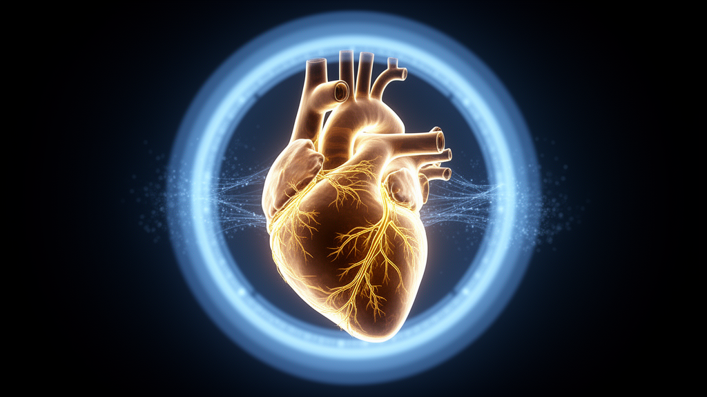
Researchers have developed a computational method that builds patient-specific three-dimensional models of the coronary arterial tree, extending from the large epicardial vessels down to microscopic branches, using data drawn from clinical myocardial perfusion PET imaging. The work, published as original research in EJNMMI Physics, addresses a longstanding gap: positron emission tomography myocardial perfusion imaging (PET-MPI) is widely used to quantify myocardial blood flow, but on its own it does not resolve the dynamic coupling between coronary flow, microvascular flow and myocardial blood volume that underlies the pathophysiology of ischemic heart disease.
The new approach adapts constrained constructive optimization, or CCO, an algorithmic technique for generating bifurcating arterial trees. In the reported implementation, the model ingested each patient’s three-dimensional myocardial geometry from static PET, voxel-wise myocardial blood flow values derived from dynamic Rb-82 PET kinetic modeling, epicardial stenosis information from fluoroscopy imaging, and a model of the epicardial coronary vessels that served as the starting point. From there, the algorithm grew bifurcating arterial trees extending from the epicardial vessels toward the endocardium, constrained by the PET-derived blood flow distribution, physiological branching morphometry and a rheological model governing flow behavior.
The method was evaluated in six patients with myocardial ischemia. According to the results, the generated vascular networks produced terminal vessels with approximate diameters of 120 micrometers, placing the smallest modeled branches at the scale of the microcirculation. Reproducibility was high across random initializations, with a mean-square-error below 0.02, meaning that repeated runs of the algorithm starting from different random conditions converged on closely matching network structures.
Anatomical plausibility was assessed against an independent benchmark. Vessel diameters in the generated trees showed excellent agreement with published porcine morphometric data, with a correlation coefficient greater than 0.99 on a log-log scale. While the comparison data come from pig heart measurements rather than human histology, the close fit suggests the algorithm reproduces established scaling relationships in vascular architecture.
Functionally, the simulated tissue-level perfusion closely matched the PET-derived values used to constrain the models. In ischemic regions, the model reproduced a reduction in flow relative to remote myocardial tissue, mirroring the pattern seen in the patients’ own imaging. A mixed-effects analysis revealed a moderate to strong correlation, r = 0.60, between the count of terminal vascular segments and segmental perfusion, indicating that structural features of the generated networks carry quantitative information about regional blood supply.
Because the generated networks support pulsatile hemodynamic simulation, the framework allows researchers to compute how blood moves through a patient’s reconstructed coronary tree under realistic cyclic driving pressures. This is the step that connects static perfusion maps to the physics of flow through branching vessels of widely varying diameter, from millimeter-scale epicardial arteries down to arterioles around 120 micrometers across.
The authors position the platform as a foundation for future in-silico derivation of invasive clinical indices, specifically coronary flow reserve and the index of microcirculatory resistance, across a spectrum of ischemic heart diseases. The study, however, reports model generation and simulation performance, not clinical validation of such derived indices against catheter measurements.
Several limitations frame the findings. The evaluation included six patients, all with myocardial ischemia, and the morphometric benchmark was porcine rather than human. The article is being shared early as a peer-reviewed, accepted manuscript carrying a permanent DOI; this version remains subject to further edits before the final Version of Record replaces it. Within those bounds, the work demonstrates that patient-specific PET perfusion data can constrain a multiscale coronary network model faithfully enough to reproduce measured perfusion patterns, offering a computational bridge between clinical imaging and detailed hemodynamic simulation.
Subject of Research: Patient-specific computational generation of coronary microvascular networks from PET myocardial perfusion imaging for hemodynamic simulation
Article Title: PET-guided adaptive CCO for patient-specific multiscale coronary arterial network generation and hemodynamic simulation
Article References: Teeraratkul, C., Mahmoudi, M., Pogosyan, A., Li, Y., Faure, S., Besson, F. L., & Nguyen, K.-L. (2026). PET-guided adaptive CCO for patient-specific multiscale coronary arterial network generation and hemodynamic simulation. EJNMMI Physics. https://doi.org/10.1186/s40658-026-00944-7
Image Credits: AI Generated
DOI: 10.1186/s40658-026-00944-7
Keywords: PET myocardial perfusion imaging, coronary arterial network, constrained constructive optimization, myocardial blood flow, hemodynamic simulation, ischemic heart disease, microcirculation, Rb-82 PET, in-silico modeling, coronary flow reserve, PET-guided, adaptive
Cite Scienmag News
Denise Maddox. (October 29, 2026). PET Scans Guide Computer-Generated Coronary Networks for Heart Blood Flow Simulation. Scienmag. https://scienmag.com/pet-scans-guide-computer-generated-coronary-networks-for-heart-blood-flow-simulation/
Denise Maddox. "PET Scans Guide Computer-Generated Coronary Networks for Heart Blood Flow Simulation." Scienmag, 29 October 2026, https://scienmag.com/pet-scans-guide-computer-generated-coronary-networks-for-heart-blood-flow-simulation/. Accessed 29 October 2026.
Denise Maddox. "PET Scans Guide Computer-Generated Coronary Networks for Heart Blood Flow Simulation." Scienmag. October 29, 2026. https://scienmag.com/pet-scans-guide-computer-generated-coronary-networks-for-heart-blood-flow-simulation/