


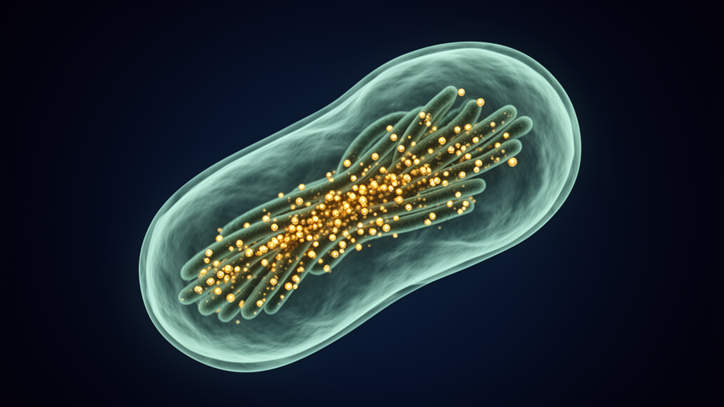
The diarrheal parasite Cryptosporidium parvum organizes its chromosomes in a way never before seen in a single-celled eukaryote, according to a new study published in PLOS Pathogens. Researchers at Washington University in St. Louis report that instead of the single, localized centromere found on each chromosome of its close relatives, C. parvum distributes hundreds of small centromere sites along all eight of its chromosomes, a configuration known as holocentricity. Until now, this architecture had been documented only in certain plants, insects, and nematodes, making the parasite the first protist in which such evidence has been described.
C. parvum is an obligate intracellular parasite and a widespread cause of diarrheal disease worldwide, posing particular danger to infants, young animals, and immunocompromised adults. Infection begins with the ingestion of infectious oocysts, after which sporozoites invade host cells and multiply through a process called merogony: three rounds of nuclear division within a single shared cytoplasm, followed by cytokinesis that produces eight infectious merozoites. Despite the medical and agricultural importance of this parasite, the mechanics of how it divides its chromosomes have remained largely uncharacterized, limited until now to basic microscopy observations.
Apicomplexan parasites, the group to which C. parvum belongs along with the malaria agent Plasmodium falciparum and the model organism Toxoplasma gondii, divide in unusual ways. They maintain the nuclear envelope throughout mitosis, keep their chromosomes minimally condensed, and typically anchor centromeres at the apical end of the nucleus while telomeres cluster at the opposite, basal pole. Centrosomes, or spindle pole bodies, are embedded in the nuclear envelope and extend microtubules that attach to centromeres to coordinate chromosome movement. In T. gondii and P. falciparum, each chromosome carries a single regional centromere, a gene-poor island of heterochromatin several to hundreds of kilobases long.
To probe centromere organization in C. parvum, the team first identified the parasite’s centromere-specific histone variant, CENH3, the defining protein component of centromeres across eukaryotes. Bioinformatic analyses, including BLASTP searches and phylogenetic reconstruction, confirmed that the gene cgd4_2030 encodes the CENH3 ortholog, distinct from two conventional histone H3 variants named H3.1 and H3.2. Using CRISPR-Cas9 gene editing, the researchers generated transgenic parasites carrying epitope-tagged versions of these histones and examined them with high-resolution confocal microscopy with Airyscan detection during the first round of merogony in human intestinal HCT-8 cell cultures.
The results were striking. While the conventional H3 variants showed the expected diffuse nuclear staining, the tagged CENH3 did not form discrete foci as it does in other apicomplexans. Instead, it stained diffusely across the apical half of the nucleus, and this pattern persisted throughout mitosis. As a control, the team examined a CENH3-tagged line of T. gondii, which displayed the punctate staining pattern characteristic of its regional centromeres. The contrast between the two parasites was sharp and consistent across different tagging strategies, including N-terminal tags, a multi-epitope spaghetti monster tag, and second copies inserted at a neutral genomic locus.
The researchers also tracked centrosomes using antibodies against centrin-1, which mark a single spot in non-dividing cells and duplicated spots during replication. All nuclei sharing a cytoplasm within a meront displayed the same centrin pattern, suggesting that nuclear division proceeds synchronously across the parasite’s shared cytoplasm, in contrast to the asynchronous divisions seen in Plasmodium schizogony. Staining for alpha-tubulin revealed microtubules forming incomplete, branched hemi-spindle structures during division, and antibodies against phosphorylated histone H3 confirmed that meront chromatin was mitotically active even though the chromosomes never visibly condense.
A second surprise concerned the telomeres. In other apicomplexans, telomeres occupy the pole opposite the centromeres, but in C. parvum a tagged version of the G-quadruplex binding protein, a marker for telomeric repeats, localized to the same apical region as CENH3. A dual-tagged parasite line expressing both fluorescently labeled proteins showed extensive overlap, quantified with an average Pearson correlation coefficient of approximately 0.90. This co-localization of centromeres and telomeres at one nuclear pole further distinguishes C. parvum from its relatives and is consistent with a fundamentally different chromosome architecture.
To map the centromeres at genomic resolution, the team adapted CUT&RUN, a technique that captures protein-bound DNA fragments after enzymatic release, using parasites expressing a heavily tagged CENH3. Validation in T. gondii reproduced previously published ChIP-seq findings, identifying a single centromere on each of that parasite’s 13 chromosomes, marked by enrichment of the heterochromatin mark H3K9me3 and depletion of the active mark H3K4me3. In C. parvum, however, the same approach revealed no dominant peaks. Instead, after optimizing nuclease digestion times, because CENH3 nucleosomes proved hypersensitive to digestion in a manner reminiscent of the holocentric nematode Caenorhabditis elegans, the researchers identified roughly 350 high-confidence CENH3 binding sites in coding regions and about 60 in intergenic regions, distributed across all eight chromosomes. These sites also correlated with H3K9me3 and lacked H3K4me3, supporting their identity as centromeric chromatin, and motif analysis revealed GA-rich repeat sequences enriched at these sites.
Notably, most of the CENH3-associated windows fell within gene coding regions, an unexpected location for centromeric DNA. Nucleotide diversity and dN/dS analyses comparing C. parvum isolates showed that these regions are highly conserved and under strong purifying selection, suggesting they are functionally important. The authors propose that the parasite may rely on numerous micro-kinetochores distributed along each chromosome, spreading the mechanical load of chromosome segregation across many small attachment points. Such a system could be advantageous for an organism that performs closed mitosis with uncondensed chromatin and must execute multiple rapid rounds of division during merogony. They also speculate that CENH3 binding in coding regions might be tolerated during mitosis, when transcription is silenced, and displaced when genes are active, though this remains to be tested.
The authors interpret their findings cautiously. They acknowledge that alternative explanations, such as asynchronous chromatid separation or cohesion fatigue, could contribute to the diffuse CENH3 pattern, and current imaging resolution cannot fully distinguish among these possibilities. The number of predicted CENH3 loci, however, far exceeds the eight regional centromeres expected under a monocentric organization. Holocentric chromosomes are thought to have evolved convergently at least 13 times across plants and animals, and the direction of centromere evolution within the Apicomplexa remains unresolved: because centromere organization has not been characterized in close outgroups such as gregarines or chromerids, C. parvum’s architecture may be either an independently derived state or a retention of an ancestral condition. Holocentricity may also confer resilience, stabilizing chromosomal fragments under the DNA-damaging stresses of the gastrointestinal environment, though it poses challenges for meiosis that well-studied holocentric species solve with specialized mechanisms such as inverted meiosis, for which the authors found no evidence in prior C. parvum crosses. Direct cytological confirmation will be difficult because the parasite’s chromosomes never condense, but the study establishes C. parvum as an intriguing new model for how chromosome architecture can diversify among eukaryotic microbes.
Subject of Research: Holocentric centromere organization in the apicomplexan parasite Cryptosporidium parvum
Article Title: Evidence for holocentric centromeres in the early branching apicomplexan parasite Cryptosporidium parvum
Article References: Kimball, A., Huang, W., Key, M., Xu, R., Funkhouser-Jones, L., & Sibley, L. D. (2026). Evidence for holocentric centromeres in the early branching apicomplexan parasite Cryptosporidium parvum. PLOS Pathogens, 22(10), e1014708. https://doi.org/10.1371/journal.ppat.1014708
Image Credits: AI Generated
DOI: 10.1371/journal.ppat.1014708
Keywords: Cryptosporidium parvum, centromere, holocentric chromosomes, CENH3, mitosis, apicomplexa, CUT&RUN, telomeres, chromosome segregation, Toxoplasma gondii, genome stability, protists
Cite Scienmag News
Juliet Wilcox. (October 26, 2026). Parasite Defies Textbook Genetics With Hundreds of Centromeres Scattered Across Its Chromosomes. Scienmag. https://scienmag.com/parasite-defies-textbook-genetics-with-hundreds-of-centromeres-scattered-across-its-chromosomes/
Juliet Wilcox. "Parasite Defies Textbook Genetics With Hundreds of Centromeres Scattered Across Its Chromosomes." Scienmag, 26 October 2026, https://scienmag.com/parasite-defies-textbook-genetics-with-hundreds-of-centromeres-scattered-across-its-chromosomes/. Accessed 26 October 2026.
Juliet Wilcox. "Parasite Defies Textbook Genetics With Hundreds of Centromeres Scattered Across Its Chromosomes." Scienmag. October 26, 2026. https://scienmag.com/parasite-defies-textbook-genetics-with-hundreds-of-centromeres-scattered-across-its-chromosomes/


