


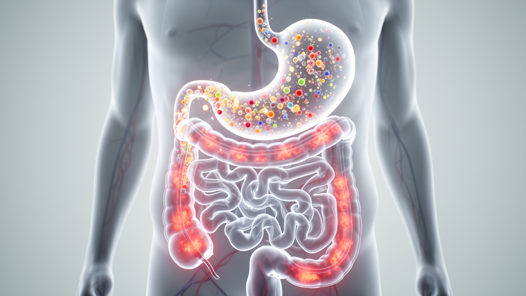
Every mechanistic claim in nutrition science travels a hidden journey before it earns its keep. A molecule in a meal is not the same molecule that reaches the bloodstream, and the molecule in the bloodstream is not necessarily the one that acts on a remote tissue. Two researchers from the United Arab Emirates argue that this gap between what a meal contains and what it is claimed to do is where much of modern mechanistic nutrition research quietly overreaches. In a conceptual analysis published in Frontiers in Nutrition, Abdulmannan Fadel of United Arab Emirates University and Yazan Ranneh of Al Ain University propose a framework designed to close that gap: the Postprandial Bioactive Exposure Axis, or PBEA.
The core problem the authors identify is compartment confusion. Mechanistic nutrition studies often begin with a food, nutrient or named bioactive and end with a claim about biological effect, skipping the exposure steps in between. Yet food-derived molecules must pass through digestion, intestinal interaction, epithelial transfer, microbial conversion and post-absorptive handling before any downstream inflammatory response can be interpreted. Each of these steps can change the chemical identity, concentration, timing and site of action of the molecules involved. A compound may be abundant in food yet poorly released from the food matrix, released but poorly absorbed, absorbed mainly after microbial transformation, or detected in blood in a form that bears little relevance to a distant tissue. When these transitions are not clearly defined, interpretation can extend beyond what the data reasonably support, particularly when causal or quasi-causal language is used about inflammation, barrier function or systemic effects.
The PBEA organizes the postprandial sequence into four operational fractions: bioaccessible, intestinally bioactive, absorbed and systemically relevant. The authors are careful to stress that these fractions are levels of exposure inference, not discrete biological compartments. The model begins with digestion-generated luminal chemistry, moves to local activity at the intestinal interface, then to net epithelial transfer into portal blood or lymph, and finally to remote tissue exposure in forms and amounts compatible with downstream signaling pathways. The term axis describes an ordered sequence of interpretation, not a validated causal pathway, and the order should not be read as a linear kinetic pathway because gastric emptying, intestinal transit, bile delivery, first-pass metabolism, lymphatic transport, tissue uptake and systemic clearance can each alter the timing, concentration and chemical form of postprandial exposure.
The first fraction, bioaccessibility, is generated by digestion rather than by the ingredient list. Standardized static and semi-dynamic INFOGEST workflows have made it easier to study how matrix structure, food processing and digestive conditions affect luminal release. Matrix effects are often large: particle size, emulsification, protein-lipid interactions, fiber entrapment and thermal processing can all alter the release and physical presentation of food-derived molecules. Structured matrices may generate early and delayed phases of luminal release as their physical structure progressively breaks down, meaning that gels, solid foods and emulsions of similar composition may produce different exposure profiles. Digestion can also create locally relevant species that were not present in the intact food, including oxidized lipid products and new peptide pools. The physical presentation matters too, since lipophilic compounds may partition into mixed micelles, dietary proteins can bind bioactives or form surface coronas around food-derived particles, and extracellular vesicles may protect encapsulated molecules during digestion. Within the PBEA, these supramolecular structures are treated as properties of the bioaccessible fraction rather than separate fractions, because they modify the form in which a compound is presented to the intestinal interface.
The second fraction captures local action at the intestinal interface, which the authors describe as a dynamic mucosal signaling site rather than a simple seal. The barrier comprises mucus, epithelial cells with their junctional complexes, and mucosal immune cells that continually survey and respond to luminal signals. Recent primary studies show that microbial activity can directly remodel gastrointestinal mucus, that tissue-resident immune cells can integrate within the intestinal epithelium, and that human intestinal organoid-on-chip systems display stimulus-dependent epithelial responses rather than passive exclusion. Luminal material can act locally without ever becoming systemically available: cytokines, dietary components, microbial metabolites and digestion products can all influence junctional behavior, epithelial metabolism, mucus properties and immune signaling at this interface. That is the logic behind the intestinally bioactive fraction, and it is why a digestion product can be bioaccessible without being intestinally bioactive, and intestinally bioactive without being absorbed.
The third and fourth fractions separate absorption from systemic relevance. Net absorption is not simply movement of material from lumen to bloodstream but a continuum of processes including apical uptake, intracellular metabolism, efflux, basolateral transport and presystemic elimination. The absorbed fraction is therefore a chemically fractionated product of transport and metabolism, not merely the bioaccessible fraction that crossed the epithelial barrier. This distinction is especially relevant for polyphenols, whose plasma and urine profiles commonly contain phase II conjugates and microbially derived catabolites rather than the parent compounds present in food. Even systemic exposure does not guarantee tissue access, since tissues protected by additional barriers such as the blood-brain barrier may never encounter a compound that appears freely in plasma. A compound may cross an epithelial model or appear in plasma yet still be present in a chemical form, concentration or time window unlikely to explain a remote biological effect.
Microbial metabolism extends the postprandial exposure sequence into the colon. For substrates that escape substantial small-intestinal absorption, colonic fermentation generates short-chain fatty acids from fibers and converts incompletely absorbed polyphenols into smaller phenolic metabolites that appear later in plasma and urine than the early small-intestinal phase. The authors offer indicative time windows: the first 0 to 4 hours after ingestion predominantly reflects upper-gastrointestinal absorption and early host metabolism, metabolite appearance from roughly 8 hours onward is more suggestive of colonic microbial transformation, and the intervening 4 to 8 hours should be considered transitional. These windows vary between meals and individuals, and human metabotype studies show that the same polyphenol-rich food can lead to different circulating metabolite profiles between people. A vascular, immune or metabolic response appearing later in the postprandial period may therefore be linked to microbial or host-microbial products rather than the parent dietary compound.
Endotoxemia serves as the paper’s flagship example of compartment-sensitive interpretation. High-fat meal studies sometimes move too quickly from postprandial lipid handling or endotoxin-related markers to broader claims about gut permeability, lipopolysaccharide translocation and inflammation. The human evidence is uneven for two reasons. First, the response appears to depend on host phenotype and postprandial lipid handling, with controlled studies reporting greater changes in endotoxin-related markers and broader inflammatory or transcriptomic responses in adults with obesity or larger postprandial triglyceride excursions. Second, the analytical problem is substantial: the Limulus amebocyte lysate assay performs poorly in plasma unless masking and recovery issues are addressed, and endotoxin recovery can be strongly affected by plasma components and protein-endotoxin interactions. Within the PBEA, lipopolysaccharide-binding protein, soluble CD14 and LAL-based endotoxin measurements can support endotoxin-related response claims when assay limitations are addressed, but they should not be treated as stand-alone evidence of increased gut permeability or direct LPS translocation. The authors also caution that widely used commercial zonulin ELISA kits have shown limited analytical specificity, and serum zonulin-family measurements have not correlated well with physiologic permeability assessed by lactulose-mannitol testing.
The model’s most practical contribution is model-to-compartment matching. Each experimental system captures only part of the postprandial sequence, so the endpoint should match the claim being made. Static and semi-dynamic digestion systems are best matched to the bioaccessible fraction. Epithelial systems such as Caco-2, Caco-2/HT29-MTX and organoid-derived monolayers support claims about intestinal bioactivity when endpoints include TEER, permeability flux, junctional proteins, oxidative stress or cytokine release, while apical-to-basolateral transfer with basolateral metabolite recovery supports narrower claims about the absorbed fraction. Fermentation models match delayed microbial transformation, and human meal studies are needed for integrated postprandial physiology. The authors propose a simple decision sequence: identify the measured compartment, define the chemical species measured, check whether sampling timing matches the expected biology, limit the claim to the highest compartment with direct supporting data, and never ascribe a systemic mechanism unless exposure, time, concentration and a biologically compatible endpoint were all measured. When a single study provides evidence for more than one PBEA fraction, claims should be limited to the highest fraction directly measured, with lower fractions used as supporting evidence.
The authors are candid about the model’s limitations. The PBEA provides no original datasets, mathematical simulations or quantitative transfer values between fractions, and should not be treated as a predictive exposure model. Its evidence base is heterogeneous, spanning digestion models, epithelial systems, fermentation experiments, metabolite profiling studies and human postprandial trials that each answer different questions and should not be treated as equivalent forms of validation. The model is likely to be most useful in research on polyphenols, lipids, microbial metabolites, barrier interpretation, endotoxin-related markers and cardiometabolic inflammation, where confusion between intake, exposure and mechanism is common. Its value, the authors conclude, lies not in defining a single pathway from meals to inflammation but in helping researchers design better studies, choose appropriate models, interpret timing more carefully and keep mechanistic claims within the evidence actually generated by postprandial nutrition studies.
Subject of Research: Nutrition Science
Article Title: From digestion to inflammatory signaling: the Postprandial Bioactive Exposure Axis as a compartment-based conceptual model for mechanistic nutrition research
Article References: Fadel, A., & Ranneh, Y. (2026). From digestion to inflammatory signaling: the Postprandial Bioactive Exposure Axis as a compartment-based conceptual model for mechanistic nutrition research. Frontiers in Nutrition, 13, Article 1859744. https://doi.org/10.3389/fnut.2026.1859744
Image Credits: AI Generated
DOI: 10.3389/fnut.2026.1859744
Keywords: postprandial metabolism, bioaccessibility, intestinal barrier, mechanistic nutrition, inflammation, gut microbiota, endotoxemia, polyphenols, bioavailability, PBEA, cardiometabolic health, exposure axis
Cite Scienmag News
Daisy Hatcher. (October 17, 2026). New Model Maps the Hidden Journey of Food Molecules From Digestion to Inflammation. Scienmag. https://scienmag.com/new-model-maps-the-hidden-journey-of-food-molecules-from-digestion-to-inflammation/
Daisy Hatcher. "New Model Maps the Hidden Journey of Food Molecules From Digestion to Inflammation." Scienmag, 17 October 2026, https://scienmag.com/new-model-maps-the-hidden-journey-of-food-molecules-from-digestion-to-inflammation/. Accessed 17 October 2026.
Daisy Hatcher. "New Model Maps the Hidden Journey of Food Molecules From Digestion to Inflammation." Scienmag. October 17, 2026. https://scienmag.com/new-model-maps-the-hidden-journey-of-food-molecules-from-digestion-to-inflammation/


