


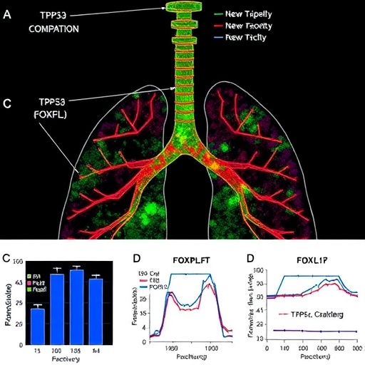
Immune checkpoint inhibitors have transformed the treatment of lung adenocarcinoma, yet they fail in nearly half of the patients who receive them, and the reasons for that failure have remained stubbornly opaque. A new computational study published in BMC Bioinformatics proposes a striking molecular explanation: a mutant form of the famous tumor suppressor p53 may physically hijack a transcription factor called FOXP3, dragging it away from the promoter of the PD-L1 gene and thereby releasing the brakes on one of cancer’s most potent immune-evasion machinery. The work, carried out by independent researcher Dev Sudersan Venkatesan of Chennai, India, weaves together six layers of computational evidence spanning clinical survival data, chromatin profiling, protein structure prediction, and molecular dynamics simulation, and it arrives at a candidate mechanism that, if confirmed experimentally, could reshape how clinicians think about immunotherapy resistance in lung cancer.
The central hypothesis is deceptively simple. FOXP3, best known as the master regulator of regulatory T cells, also acts inside tumor cells themselves, where it binds directly to the promoter of CD274, the gene encoding programmed death-ligand 1, or PD-L1, and represses its transcription. When FOXP3 can reach the promoter, PD-L1 expression is held in check. Venkatesan hypothesized that gain-of-function mutant p53, the kind of p53 mutation that does not merely disable the protein but arms it with new oncogenic activities, might bind FOXP3 directly and sequester it away from DNA. Stripped of its transcriptional regulator, the CD274 promoter would be left unguarded, allowing PD-L1 to accumulate on the tumor cell surface and blunt the effect of PD-1/PD-L1 blockade antibodies.
To test this idea computationally, the study first turned to clinical reality. Across three independent lung adenocarcinoma cohorts, the Cancer Genome Atlas PanCancer Atlas with 510 patients, the Singapore-based OncoSG cohort with 181 patients, and the Clinical Proteomic Tumor Analysis Consortium cohort with 110 patients, for a combined total of 670 individuals, the author asked whether disruption of the FOXP3-PD-L1 axis predicted patient outcomes. It did, and strongly. Patients whose tumors showed a broken FOXP3-PD-L1 relationship had significantly inferior overall survival, with a log-rank p-value of 6 × 10⁻⁴ and a hazard ratio of 1.48, meaning a 48 percent increase in the risk of death, with a 95 percent confidence interval running from 1.12 to 1.95. The association held across cohorts that differ in ancestry, treatment patterns, and genomic profiling methods, lending epidemiological weight to what is otherwise a purely theoretical construct at this stage.
The next layer of evidence concerned the DNA itself. Using the FIMO motif-scanning tool with the JASPAR 2024 position weight matrix for FOXP3, the study identified eight candidate FOXP3 consensus binding motifs, each matching the sequence GTAAACA, along the CD274 promoter, a result significant at a p-value of 7.93 × 10⁻⁵. These are the positions where FOXP3 would be expected to dock if it were free to do so. Importantly, the author is explicit that these candidate sites await confirmation by chromatin immunoprecipitation sequencing, the gold-standard experimental technique for mapping where a transcription factor actually sits on the genome. The motifs establish plausibility, not proof.
Chromatin accessibility added a crucial element of biological specificity. Analysis of ATAC-seq data, which measures how open and transcriptionally permissive regions of the genome are, revealed that the CD274 promoter is restricted, or physically less accessible, in lung adenocarcinoma but not in head and neck squamous cell carcinoma. This lineage-specific pattern matters because it begins to explain a long-standing puzzle: why PD-L1 dysregulation tied to p53 mutation appears to behave differently in different tumor types. A mechanism that depends on chromatin context rather than on mutation status alone could account for the fact that the FOXP3-checkpoint uncoupling observed in a companion pan-cancer analysis was present in 67 percent of adenocarcinomas but in exactly zero percent of non-adenocarcinoma tumors among 4,205 samples drawn from eight TCGA cohorts.
The structural heart of the study lies in its protein modeling. Using AlphaFold 3, the deep-learning system from Google DeepMind that predicts the structures of protein complexes, the author modeled a heterodimer between mutant p53 and FOXP3. The predicted interface showed confident local geometry, with predicted local distance difference test scores above 70 at the contact region, indicating that the model considers the physical association well supported at the residue level. Predicted aligned error analysis, which estimates the reliability of relative domain placements, further supported the plausibility of a stable complex rather than a chance collision of two unrelated proteins.
Structure alone, however, says nothing about stability in the crowded, thermal environment of a living cell. To address that, the study turned to classical molecular dynamics. The predicted complex was solvated in a TIP3P water model and simulated with the AMBER ff19SB force field under physiological conditions: an isobaric-isothermal ensemble at 310 kelvin, a salt concentration of 0.15 molar sodium chloride, run for 2 nanoseconds on an NVIDIA A100 graphics processing unit. Over the course of the simulation, the complex underwent progressive compaction, with the radius of gyration contracting from 46.7 to 43.9 angstroms, a sign that the two chains were folding into one another rather than drifting apart. Correlated motion analysis showed significant inter-chain coordination, meaning the two proteins moved as a single mechanical unit, and root-mean-square fluctuation analysis of the interface residues between positions 150 and 300 revealed rigid geometry in the 1 to 2 angstrom range. The estimated interaction energy of the complex was −55.33 kilocalories per mole, a substantially favorable figure consistent with a stable physical association.
The final strand of evidence, drawn from a previously published preprint cited within the study, connects the structural story back to gene expression in real tumors. Among 517 TCGA lung adenocarcinoma samples, mutant p53 status was associated with significant upregulation of CD274, with a log2 fold change of 0.53 and an adjusted p-value below 0.0001, while FOXP3 expression itself was untouched, with a log2 fold change of just 0.014 and an adjusted p-value of 0.889. That dissociation is exactly what the sequestration model predicts: if mutant p53 were simply reducing FOXP3 production, FOXP3 mRNA would fall alongside rising PD-L1. Instead, FOXP3 remains present but appears functionally sidelined, unable to reach its target promoter and do its repressive work.
Taken together, the six layers of evidence form a coherent, if still provisional, narrative. Gain-of-function mutant p53, one of the most common molecular lesions in lung adenocarcinoma, may act as a molecular decoy for FOXP3, occupying it in the nucleoplasm and preventing promoter occupancy at eight candidate sites on CD274. The consequence is unrestrained PD-L1 expression, impaired immune surveillance, and measurably worse survival across 670 patients. Because the effect appears confined to adenocarcinoma lineages, the model also offers a testable explanation for why checkpoint therapy outcomes differ so markedly between lung adenocarcinoma and squamous histologies.
The author and the field alike are careful to emphasize what the study does not yet show. Computational prediction, however multi-layered, is not experimental demonstration. The critical missing pieces are co-immunoprecipitation experiments to confirm that mutant p53 and FOXP3 physically associate in cells, ChIP-seq to confirm FOXP3 occupancy loss at the CD274 promoter, and promoter reporter assays to show that FOXP3-mediated repression of CD274 is relieved by mutant p53 in a dose-dependent fashion. Until those experiments are done, the mutp53-FOXP3 interaction remains a candidate mechanism, albeit one supported by an unusually broad convergence of independent data types.
If validation succeeds, the therapeutic implications could be considerable. The mutp53-FOXP3 interface would become a high-priority drug target, and restoring FOXP3 access to the CD274 promoter, whether by disrupting the sequestration interaction or by designing combination regimens around it, could convert a subset of immunotherapy-resistant lung adenocarcinoma patients into responders. The study also underscores a broader lesson for computational oncology: when survival statistics, chromatin accessibility, structural prediction, and molecular dynamics all point in the same direction, even a single-author, unfunded effort conducted on cloud computing resources can generate hypotheses worthy of the laboratory’s full attention. For now, the mutant p53-FOXP3 axis stands as one of the most intriguing candidates yet proposed for explaining why so many lung cancer patients do not benefit from the immunotherapy revolution.
Cite Scienmag News
Nathaniel Bowman. (September 8, 2026). Mutant TP53 traps FOXP3, disrupting PD-L1 control and fueling immune evasion in lung cancer. Scienmag. https://scienmag.com/mutant-tp53-traps-foxp3-disrupting-pd-l1-control-and-fueling-immune-evasion-in-lung-cancer/
Nathaniel Bowman. "Mutant TP53 traps FOXP3, disrupting PD-L1 control and fueling immune evasion in lung cancer." Scienmag, 8 September 2026, https://scienmag.com/mutant-tp53-traps-foxp3-disrupting-pd-l1-control-and-fueling-immune-evasion-in-lung-cancer/. Accessed 8 September 2026.
Nathaniel Bowman. "Mutant TP53 traps FOXP3, disrupting PD-L1 control and fueling immune evasion in lung cancer." Scienmag. September 8, 2026. https://scienmag.com/mutant-tp53-traps-foxp3-disrupting-pd-l1-control-and-fueling-immune-evasion-in-lung-cancer/


