


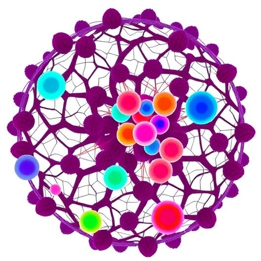
In a comprehensive new review published in Molecular Biology Reports, researchers from Hangzhou Medical College and Zhejiang Provincial People’s Hospital have assembled one of the most detailed syntheses to date of how macrophages—the immune system’s most versatile scavenger cells—drive, sustain, and sometimes reverse fibrosis in the lung, liver, kidney, and heart. Their central argument is provocative: the textbook M1/M2 polarization model that has dominated immunology teaching for decades is no longer adequate to explain what actually happens inside fibrotic organs, and the single-cell omics revolution has exposed a degree of macrophage diversity that far exceeds this binary framework.
The stakes of the review are considerable. Organ fibrosis, the end result of chronically dysregulated tissue repair, is estimated to account for roughly 45 percent of all deaths worldwide, spanning conditions as diverse as idiopathic pulmonary fibrosis, cirrhosis, chronic kidney disease, and post-infarction cardiac scarring. Despite its enormous clinical burden, there are currently no treatments that reliably reverse established fibrosis; existing therapies such as nintedanib, pirfenidone, and resmetirom slow progression at best. By mapping the immune landscape of fibrosis at single-cell and single-cell-location resolution, the review’s authors argue that macrophages represent the most promising lever for precision interventions—and that the field’s failure to exploit their heterogeneity has been a key reason anti-fibrotic drug development has repeatedly stalled.
The technical foundation for this reappraisal is the maturation of single-cell RNA sequencing, single-nucleus RNA sequencing, and spatial transcriptomics. These platforms allow investigators to profile thousands of individual cells from fibrotic tissue, cluster them by their transcriptional identities, and then place those identities back into their anatomical context within the scarred organ. What these tools have revealed is that macrophages within a single fibrotic organ are not a uniform population responding uniformly to inflammation. Instead, they occupy a multidimensional landscape defined by at least four axes: their cellular origin, the stage of disease at which they are sampled, the spatial microenvironment they inhabit, and their ultimate functional output, whether that output is collagen deposition, matrix degradation, or efferocytosis—the clearance of dead cells.
The origin axis is particularly consequential. Tissue-resident macrophages in organs such as the lung, liver, and brain are embryonically derived, seeding tissues during development from yolk sac and fetal liver progenitors and thereafter maintaining themselves locally through self-renewal. In the liver, these resident cells are the Kupffer cells, which patrol the sinusoids and perform surveillance functions honed over a lifetime. By contrast, when injury strikes, bone marrow-derived monocytes flood into the damaged organ in response to chemokine gradients—chiefly the CCL2/CCR2 axis—and differentiate into monocyte-derived macrophages with fundamentally different transcriptional programs. Fate-mapping studies using markers such as Ms4a3 have allowed researchers to distinguish these lineages with precision, and the distinction matters therapeutically: in the lung, monocyte-derived alveolar macrophages have been shown to drive fibrosis and persist in tissue over the lifespan of the animal, whereas resident alveolar macrophages generally perform homeostatic and restorative functions.
Nowhere is the new heterogeneity framework more vividly illustrated than in the identification of SPP1-expressing macrophages. Secreted Phosphoprotein 1, also known as osteopontin, marks a subset of stroma-associated macrophages that the review identifies as a conserved pro-fibrotic end-state across all four organs examined. In the lung, proliferating SPP1/MERTK-expressing macrophages have been documented in idiopathic pulmonary fibrosis. In the kidney, platelet-instructed SPP1-positive macrophages drive myofibroblast activation in a CXCL4-dependent manner. In the heart, spatial multi-omic maps of human myocardial infarction have localized these cells to the infarct border zone, where they sit alongside activated fibroblasts in a self-reinforcing signaling loop. The review’s authors propose that SPP1-positive macrophages, because they recur in fibrotic niches across organ boundaries, could serve as a candidate cross-organ therapeutic axis—meaning a drug developed against this cell type in one organ might conceivably be repurposed for fibrotic diseases elsewhere in the body.
Spatial transcriptomics has been essential to identifying these conserved niches because it reveals not just which cells are present, but where they are relative to their targets. Scar-associated macrophages, for example, are not scattered randomly through fibrotic tissue; they cluster adjacent to myofibroblasts, the collagen-producing workhorses of fibrosis, in spatially restricted niches sustained by growth factors such as macrophage colony-stimulating factor. In the heart, CCR2-positive and CCR2-negative resident macrophages perform distinct roles in orchestrating monocyte recruitment after myocardial injury, and their positioning within the infarct zone versus the border zone correlates with different outcomes for tissue remodeling. The spatial dimension transforms macrophage biology from a cell-autonomous story into an ecological one: macrophages behave differently depending on which neighbors they are talking to.
The review also devotes substantial attention to the metabolic reprogramming that underlies macrophage fate decisions—a dimension of immunology that has accelerated dramatically since the recognition that activated immune cells undergo profound shifts in fuel preference. Classically activated pro-inflammatory macrophages rely on aerobic glycolysis, a Warburg-like metabolic state that prioritizes rapid ATP generation and biosynthetic intermediates over mitochondrial efficiency. The glycolysis–PKM2 axis, in which Pyruvate Kinase Isozyme Type M2 acts as both a metabolic enzyme and a transcriptional co-activator of HIF-1α-driven inflammatory genes, has emerged as a central switch in this process. In the kidney, myeloid PFKFB3-mediated glycolysis has been shown to promote fibrosis, and pharmacological inhibition of PFKFB3 attenuates disease in experimental models. In the heart, temporal changes in glucose metabolism reflect polarization shifts in resident and monocyte-derived macrophages after myocardial infarction, offering a metabolic readout of the transition from inflammatory to reparative phases.
Metabolism does not merely fuel macrophages; it determines what they become. The review highlights how succinate, an intermediate of the tricarboxylic acid cycle, functions as a signaling molecule through its receptor SUCNR1 to induce profibrotic M2-like macrophages in the kidney, yet exerts protective effects on hepatocytes in the context of non-alcoholic fatty liver disease. This context-dependence—where the same metabolite promotes pathology in one organ and protects in another—illustrates why the review’s authors caution against oversimplified cross-organ generalizations even as they identify conserved subsets like SPP1-positive macrophages. Similarly, the c-Rel transcription factor has been shown to orchestrate energy-dependent reprogramming in both epithelial cells and macrophages during fibrosis, linking mitochondrial metabolism to the epigenetic regulation of inflammatory gene expression.
On the therapeutic front, the review surveys an expanding toolkit for manipulating macrophages in fibrotic disease. Rather than depleting all macrophages—a strategy that risks impairing essential homeostatic and reparative functions—emerging approaches aim for precision. Selective depletion of profibrotic macrophages using bioactivated in-vivo self-assembly peptides has been shown to ameliorate kidney fibrosis in experimental models while sparing beneficial populations. Therapeutic silencing of Spp1 specifically within TREM2-positive cardiac macrophages has been demonstrated to suppress atrial fibrillation. Nanoengineered immunosuppressive therapeutics, including dendrimer-graphite nanoparticles and endogenous cell-targeting nanoplatforms, are being explored for their ability to modulate the balance of macrophage phenotypes in situ rather than eliminating cells outright. Reprogramming strategies—shifting macrophage polarization away from profibrotic states using agents such as the FXR agonist resmetirom, which recently completed a Phase 3 trial in metabolic dysfunction-associated steatohepatitis—represent another layer of intervention that exploits metabolic vulnerabilities.
Chemokine axis blockade remains a parallel strategy. The CCR2/CCR5 dual inhibitor cenicriviroc, originally investigated for its ability to reduce inflammatory monocyte recruitment to the liver in steatohepatitis, has been re-examined in the context of COVID-19-associated lung injury, while inhibitors targeting PSMP/MSMP through CCR2 represent novel antifibrotic targets identified in hepatology. The review also underscores the significance of macrophage-to-myofibroblast transition, or MMT, in which macrophages themselves acquire collagen-producing characteristics—a phenomenon documented in renal fibrosis via the neural transcription factor Pou4f1 and in kidney disease through myofibroblast-derived exosomal signaling. Blocking these transdifferentiation pathways, rather than simply modulating polarization, adds a third dimension to the therapeutic landscape.
The authors argue that understanding macrophage heterogeneity as a multidimensional phenomenon—shaped simultaneously by origin, disease stage, spatial context, and metabolic state—will be necessary to design interventions that can navigate this complexity rather than being defeated by it. They highlight that organ-specific macrophage biology remains non-uniform despite the identification of conserved subsets, and that therapeutic strategies calibrated to a single axis of heterogeneity risk incompletely modulating disease. In liver fibrosis, for example, splenocyte-derived macrophages traveling through a spleen–liver axis exacerbate scarring independently of bone marrow-derived recruitment, adding yet another anatomical layer to macrophage origins that must be considered. In lung fibrosis, interstitial macrophages and alveolar macrophages represent distinct compartments with distinct functional roles, and treatments must account for both. In cardiac fibrosis, macrophage-derived CCL24 signaling through fibroblast CCR3 has been identified as a driver of pathological remodeling, while MMP-12-producing Ly6C-low macrophages paradoxically extend post-infarction survival by preventing neutrophil influx—underscoring that macrophages cannot be uniformly classified as friend or foe.
What the review ultimately advances is a conceptual reframing. Fibrosis research has historically sought a single target or a single pathway that could be inhibited across all patients. The single-cell omics era, as synthesized here, argues instead for precision strategies tailored to the specific macrophage subtypes and microenvironmental niches operating in a given patient’s disease at a given stage. The identification of SPP1-positive macrophages as a conserved, targetable, pro-fibrotic end-state across lung, liver, kidney, and heart offers one candidate for such a strategy, while the growing catalog of metabolic checkpoints—PKM2, PFKFB3, SUCNR1, and c-Rel—provides pharmacological handles for shifting macrophage behavior without destroying these essential cells. Whether this framework can be translated into clinical interventions that meaningfully reverse, rather than merely slow, organ fibrosis remains the defining challenge of the next decade of fibrosis research, but the review makes clear that the macrophage, in all its staggering diversity, is now the center of that effort.
Cite Scienmag News
Avery Chandler. (September 8, 2026). Single-cell omics reveal macrophage diversity in organ fibrosis. Scienmag. https://scienmag.com/single-cell-omics-reveal-macrophage-diversity-in-organ-fibrosis/
Avery Chandler. "Single-cell omics reveal macrophage diversity in organ fibrosis." Scienmag, 8 September 2026, https://scienmag.com/single-cell-omics-reveal-macrophage-diversity-in-organ-fibrosis/. Accessed 8 September 2026.
Avery Chandler. "Single-cell omics reveal macrophage diversity in organ fibrosis." Scienmag. September 8, 2026. https://scienmag.com/single-cell-omics-reveal-macrophage-diversity-in-organ-fibrosis/


