


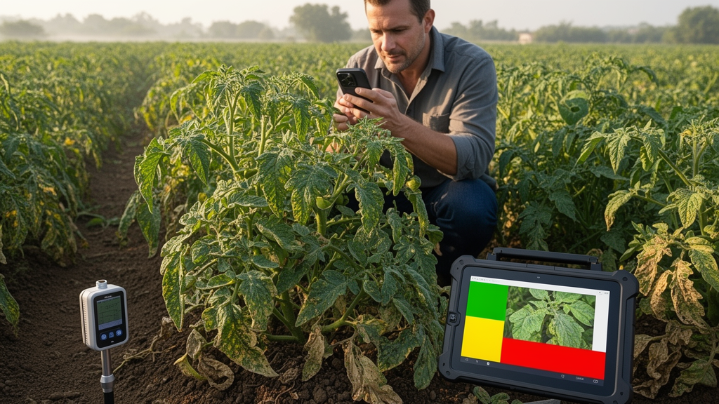
A multimodal artificial-intelligence framework called AgriMamba can assess plant disease severity by combining field images with descriptions of pathological symptoms, according to a peer-reviewed study published online on August 5, 2026 in The Crop Journal. The system first locates the leaf that needs analysis, then identifies diseased regions and calculates the proportion of the leaf covered by lesions to assign a quantitative severity grade. Researchers developed and tested the framework on a multimodal plant-disease grading dataset containing 10,350 samples from 16 crop species, reporting competitive performance against several representative deep-learning methods.
The work addresses a practical distinction between recognizing that a plant is diseased and measuring how seriously it is affected. Disease severity can indicate the extent of crop damage and may help guide disease-control strategies, pesticide use and assessment of yield losses. Image-based automation offers a way to make such assessments more efficient and objective, but field photographs present substantial visual challenges. Soil, branches, neighboring leaves, shadows and uneven illumination can surround the plant of interest, while overlapping foliage may obscure the target leaf. Small lesions may also cover only a tiny fraction of the leaf, and diseased tissue can resemble healthy tissue in color and texture, making subtle boundaries difficult for conventional vision models to distinguish.
AgriMamba was designed by a team led by Guizhou University professor Qi Wang to overcome these complications through a coarse-to-fine analytical sequence: target-leaf localization, fine-grained lesion segmentation and quantitative severity grading. Rather than asking a single model to inspect an entire cluttered field image and immediately return a severity class, the framework decomposes the task into stages. A localization-aware Mamba leaf segmenter identifies the relevant leaf and suppresses irrelevant background information. A text-guided Mamba lesion segmenter then combines visual features with pathological descriptions to locate diseased areas more precisely. The final stage measures the lesion area relative to the target leaf and maps that ratio to the corresponding disease-severity grade.
The textual component functions as an additional set of diagnostic clues. Agricultural experts do not assess a diseased leaf solely by its overall appearance; they may consider whether lesions are yellow or brown, circular or irregular, sharply or diffusely bordered, and concentrated or scattered across the leaf surface. The framework incorporates similar descriptions into visual analysis, giving the model explicit symptom characteristics that can help distinguish visually similar healthy and diseased tissues. The approach therefore uses two forms of information within one pipeline: the image supplies spatial and visual evidence, while the text specifies the pathological features that should guide attention.
The research team also examined which regions influenced the model’s decisions through visualization experiments. During the first stage, the model’s principal responses remained concentrated on the complete target leaf even when images included branches, soil, other leaves and natural background features. After the target leaf had been identified, the regions of attention in the second stage shifted progressively toward abnormal colors, changes in texture and specific lesion positions on the leaf surface. The researchers described this progression as the model narrowing its analytical focus from the whole leaf to the areas where disease appeared. These observations were consistent with the intended behavior of the staged framework, although the study summary does not provide numerical values for the attention analyses or indicate how they were statistically validated.
According to Wang, AgriMamba showed competitive overall performance when compared with multiple representative deep-learning approaches for plant-disease severity grading. The framework also maintained stable recognition performance in test situations involving small lesions, scattered lesions, partial occlusion and visually confusing backgrounds. These conditions represent important sources of difficulty in agricultural imaging because they can cause an automated system to select the wrong leaf, overlook limited areas of damaged tissue or confuse background features with symptoms. The reported stability therefore concerns recognition across these challenging visual scenarios; the source does not provide the number of test samples, severity-class distribution, comparison metrics, confidence intervals or results from an independent field trial.
Lead author Qinglong Wu said the key design choice was to replace direct assessment of a complex field image with a sequence of interpretable operations: find the leaf requiring analysis, identify its lesions, and measure the fraction affected by disease. This structure aligns the system’s output with the physical quantity used for grading. It does not rely only on an opaque image-level label, but bases the assigned grade on lesion area as a proportion of the target leaf. The framework was evaluated as an imaging-analysis method, and the researchers reported it as a peer-reviewed publication in The Crop Journal. The supplied study information does not describe deployment across a particular season, geographic region or commercial production system.
There are also important limits to the current evidence. The dataset included 10,350 multimodal grading samples drawn from 16 crop species, but the news release does not state how many images or texts contributed to each sample, how the samples were divided among training, validation and test sets, or whether images from the same plants or conditions appeared in more than one split. It likewise does not report performance separately by crop, disease, severity level or field environment. Although comparisons with several deep-learning methods were described as competitive, the release does not give the names of all comparison models, their implementation details or the numerical size of any performance advantage. Nor does it establish that the framework improves crop yields, reduces pesticide use or leads to better disease-control decisions in practice.
The authors nevertheless argue that pathology-aware, multimodal analysis could provide a useful technical approach for automated disease assessment in complex environments. Wu said that further improvements in computational efficiency and practical field deployment may allow similar methods to support automated crop scouting and smart agricultural management. Those possibilities remain future applications rather than demonstrated outcomes of the study. The strongest supported conclusion is narrower: in the researchers’ experiments, AgriMamba used staged leaf localization, text-guided lesion segmentation and area-based grading to evaluate disease severity, achieved competitive results against representative deep-learning methods, and showed stable recognition under several difficult visual conditions. The study’s DOI is 10.1016/j.cj.2026.07.003, and the authors declared no competing financial or personal interests that could have influenced the work.
Subject of Research: A multimodal imaging framework for quantitative plant disease severity grading in crop field images
Article Title: Exploring the mysteries of plant disease severity through multimodal learning
Article References: Exploring the mysteries of plant disease severity through multimodal learning. Original publication (Bibliographic details unavailable.)
Image Credits: AI Generated
DOI: Not provided
Keywords: AgriMamba, plant disease severity, multimodal learning, Mamba models, lesion segmentation, crop pathology, computer vision, agricultural imaging, disease grading, Exploring, mysteries, plant
Cite Scienmag News
Alan Morgan. (October 20, 2026). Multimodal model grades plant disease severity from field images. Scienmag. https://scienmag.com/multimodal-model-grades-plant-disease-severity-from-field-images/
Alan Morgan. "Multimodal model grades plant disease severity from field images." Scienmag, 20 October 2026, https://scienmag.com/multimodal-model-grades-plant-disease-severity-from-field-images/. Accessed 20 October 2026.
Alan Morgan. "Multimodal model grades plant disease severity from field images." Scienmag. October 20, 2026. https://scienmag.com/multimodal-model-grades-plant-disease-severity-from-field-images/


