


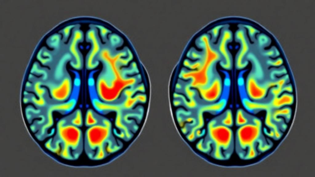
When a brain metastasis grows after stereotactic radiosurgery, oncologists face one of the most consequential questions in neuro-oncology: is the expanding mass a returning cancer, or is it radiation necrosis, a delayed injury in which the treatment itself has damaged the brain? The two can look nearly identical on conventional MRI, yet the answers demand opposite paths—one calls for systemic therapy or another round of targeted radiation, the other for corticosteroids or observation, sometimes with a risky biopsy in between. A new study published in the Journal of Neuro-Oncology now reports that a computational technique called tumor habitat analysis, applied to standard MRI sequences, can separate these entities with meaningful accuracy in a cohort where every single diagnosis was confirmed under the microscope.
The research, led by Ji Eun Park of Johns Hopkins University and Asan Medical Center together with Jingwen Yao and Benjamin M. Ellingson of the University of California, Los Angeles, set out to do what most imaging biomarker studies avoid: validate a previously published algorithm against histopathological ground truth in a fully independent cohort. The team assembled 104 patients treated between December 2011 and February 2025 at a US tertiary center, each with a contrast-enhancing brain lesion of at least one cubic centimeter and subsequent surgical or biopsy confirmation. Sixty-eight lesions proved to be recurrent metastatic tumor; thirty-six were radiation necrosis. Crucially, none of these patients, lesions, or even scanner hardware overlapped with the Asian cohort on which the habitat model had originally been built, making this a genuine test of generalizability rather than an exercise in fitting data to itself.
The underlying idea of habitat analysis is elegant. Just as ecologists divide a landscape into distinct habitats, the technique divides a tumor into biologically meaningful subregions by clustering voxels—the tiny three-dimensional pixels of an MRI volume—that share similar signal characteristics. Instead of drawing a single threshold on one image sequence and hoping it separates tumor from dead tissue, the method combines multiple sequences simultaneously. Structural habitats were derived from normalized contrast-enhanced T1-weighted and T2-weighted images, yielding three categories: enhancing tissue, solid low-enhancing tissue, and nonviable tissue. Physiologic habitats came from apparent diffusion coefficient maps, which reflect cellular density through water diffusion, and normalized cerebral blood volume maps from dynamic susceptibility contrast perfusion, yielding hypervascular, hypovascular cellular, and nonviable compartments.
Technical rigor underpinned the pipeline. Contrast-enhancing lesions were segmented using deep-learning algorithms, with manual correction where necessary. Perfusion data were motion-corrected and adjusted for the bidirectional leakage of contrast agent across a disrupted blood-brain barrier, a notorious source of error in cerebral blood volume estimation. Signal intensities were normalized against normal-appearing white matter to mitigate differences between scanners and field strengths, which ranged from 1.5 to 3 Tesla. All images were then co-registered to isotropic one-millimeter resolution. Most importantly, the k-means clustering centroids and decision boundaries frozen into the original model were applied to the new data without any retraining or refitting—a fixed yardstick carried across continents, institutions, and years.
The results showed a consistent biological signature. Recurrent tumors had larger contrast-enhancing volumes (12.2 versus 6.0 milliliters on average) and harbored a greater proportion of solid low-enhancing tissue—regions that appear dark on both T2 and contrast-enhanced T1 images, a pattern long recognized qualitatively as T1/T2 mismatch but never reliably quantified. Tumors also contained a larger hypervascular fraction, reflecting the vigorous neovasculature that feeding cancers demand, and a smaller fraction of nonviable tissue on both structural and physiologic maps. Radiation necrosis, by contrast, was dominated by nonviable habitat: on structural MRI, dead tissue made up 67.9 percent of the average necrotic lesion versus 50.2 percent of recurrent tumors, a difference that was statistically robust.
Not every habitat carried diagnostic weight. The hypovascular cellular compartment, though larger in absolute volume within tumors, proved uninformative when expressed as a fraction of the lesion. The authors attribute this to confounding biology: apparent diffusion coefficient values vary substantially across metastatic histologies—breast cancer metastases differ by receptor status, lung cancer metastases by subtype—and radiation necrosis itself lowers diffusion values through radiation-induced ischemia. This nuance matters, because a prior longitudinal study had found that growth of the hypovascular cellular habitat over time predicted future recurrence, suggesting that change over serial scans may rescue this parameter where a single snapshot cannot.
The centerpiece of the study was a composite habitat score. The researchers selected diagnostic thresholds within the current cohort—a solid low-enhancing fraction above 24.4 percent, a structural nonviable fraction at or below 57.1 percent, a hypervascular fraction above 1.3 percent, and a physiologic nonviable fraction at or below 21.1 percent—and assigned each lesion one point per threshold crossed. The combined structural and physiologic score achieved an area under the receiver operating characteristic curve of 0.80, with a sensitivity of 89.7 percent and a specificity of 58.3 percent, significantly outperforming the structural score alone. The physiologic score on its own reached an area of 0.75, and a single habitat—the hypervascular fraction—achieved an area of 0.82, comparable to meta-analytic estimates for perfusion MRI that pooled a decade of smaller, largely unvalidated studies.
How does this stack up against the alternatives? MR spectroscopy can reach sensitivities and specificities above 90 percent for choline-based ratios, but with substantial heterogeneity across studies and limited spatial information. Amino acid PET using tracers such as fluorine-18-fluoroethyl-tyrosine achieves pooled sensitivity and specificity of roughly 82 and 84 percent, but requires a cyclotron-produced radiotracer, an additional imaging session, and delivers lower spatial resolution. Habitat analysis, by contrast, extracts its answer from structural, diffusion, and perfusion sequences that are already embedded in routine brain tumor protocols, adds no radiation exposure, and—critically—maps where within a lesion the viable tumor sits. That spatial dimension could directly guide repeat radiosurgery or surgical planning by identifying the subregion most likely to harbor active disease.
The authors are candid about the limits. Specificity of 58.3 percent means the score flags far more lesions as suspicious than truly harbor tumor, so it cannot stand alone as the basis for invasive decisions; it must be read alongside serial imaging, clinical course, and multidisciplinary judgment. Multivariable modeling was impossible at this sample size, very small lesions may destabilize volume-fraction estimates, and normalization to white matter cannot erase every interscanner difference. Whether habitat analysis adds value beyond an experienced neuroradiologist’s read also remains untested. Yet the study’s central achievement stands: a model frozen on one continent transferred, unchanged, to an independent pathology-confirmed cohort on another, and still separated living cancer from treatment-wounded brain. For the growing population of patients living with irradiated brain metastases, that is a meaningful step toward answering the question that decides their next treatment—without a scalpel.
Subject of Research: MRI-based tumor habitat analysis for differentiating tumor recurrence from radiation necrosis in brain metastases after stereotactic radiosurgery
Article Title: Pathology-validated structural and physiologic habitat imaging for differentiating radiation necrosis from tumor recurrence in brain metastases
Article References: Park, J. E., Shao, G., Baisiwala, S., Kim, N., Tan, A., Sanvito, F., Liang, A., Wang, Z., Nocera, G., Raymond, C., Le, V. L., Kim, H. S., Salamon, N., Pope, W. B., Kim, W., Ellingson, B. M., & Yao, J. (2026). Pathology-validated structural and physiologic habitat imaging for differentiating radiation necrosis from tumor recurrence in brain metastases. Journal of Neuro-Oncology, 179(2), Article 61. https://doi.org/10.1007/s11060-026-05761-7
Image Credits: AI Generated
DOI: 10.1007/s11060-026-05761-7
Keywords: brain metastases, radiation necrosis, stereotactic radiosurgery, tumor habitat analysis, MRI, perfusion imaging, cerebral blood volume, apparent diffusion coefficient, pathology validation, machine learning, neuro-oncology, diagnostic imaging
Cite Scienmag News
Nathaniel Bowman. (October 4, 2026). MRI Habitat Mapping Passes Its Toughest Test: Telling Tumor Recurrence From Radiation Necrosis. Scienmag. https://scienmag.com/mri-habitat-mapping-passes-its-toughest-test-telling-tumor-recurrence-from-radiation-necrosis/
Nathaniel Bowman. "MRI Habitat Mapping Passes Its Toughest Test: Telling Tumor Recurrence From Radiation Necrosis." Scienmag, 4 October 2026, https://scienmag.com/mri-habitat-mapping-passes-its-toughest-test-telling-tumor-recurrence-from-radiation-necrosis/. Accessed 17 October 2026.
Nathaniel Bowman. "MRI Habitat Mapping Passes Its Toughest Test: Telling Tumor Recurrence From Radiation Necrosis." Scienmag. October 4, 2026. https://scienmag.com/mri-habitat-mapping-passes-its-toughest-test-telling-tumor-recurrence-from-radiation-necrosis/


