


In a groundbreaking study poised to reshape our understanding of neurodegeneration in Parkinson’s disease, researchers Kim, Cash, Martins, and colleagues have harnessed the power of multiparametric MRI combined with cutting-edge imaging transcriptomics to uncover the intricate molecular and cellular mechanisms underlying experimental Parkinsonism. Published in the esteemed journal npj Parkinsons Dis. in 2026, this investigation not only advances the frontier of neuroimaging but also bridges the crucial gap between molecular pathology and functional brain alterations, offering new vistas for early diagnosis and therapeutic interventions.
Parkinson’s disease (PD) has long challenged scientists and clinicians alike due to its complex pathology that involves a cascade of cellular events leading to the progressive loss of dopaminergic neurons in the substantia nigra. Despite extensive research, the precise molecular correlates of neurodegeneration have remained elusive, impeding the development of disease-modifying therapies. Employing a sophisticated multiparametric MRI approach, the research team achieved high-resolution, non-invasive visualization of brain tissue microstructure and functional changes in models of experimental Parkinsonism, setting a new benchmark for in vivo characterization.
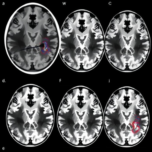
The study utilized multiple MRI parameters including diffusion tensor imaging (DTI), susceptibility-weighted imaging (SWI), and functional MRI (fMRI) to capture complementary features of brain pathology. DTI provided insights into white matter integrity by measuring fractional anisotropy, while SWI allowed for the detection of abnormal iron deposition—a hallmark of Parkinsonian neurodegeneration. fMRI further delineated alterations in resting-state connectivity that allude to disrupted neural circuits. This multipronged imaging strategy enabled a holistic view of the structural and functional deterioration unachievable through traditional single-parameter MRI scans.
However, where this research truly breaks new ground is the integration of imaging transcriptomics—an innovative technique that spatially maps gene expression patterns within brain regions identified as abnormal by MRI. By extracting and sequencing RNA from microdissected brain areas corresponding to MRI signal changes, the team delineated gene networks modulated during neurodegeneration. This dual-layer approach empowered the identification of candidate molecular pathways driving cell death and neuroinflammation, some of which had never before been linked to Parkinsonism.
Among the notable molecular findings was the heightened expression of genes implicated in microglial activation and oxidative stress responses, bolstering the increasingly supported hypothesis that neuroinflammation contributes significantly to Parkinson’s progression. Additionally, dysregulation of pathways associated with mitochondrial function and protein aggregation surfaced prominently, corroborating previous pathological observations yet now mapped precisely onto neuroimaging data. This molecular validation via imaging transcriptomics provides compelling evidence that these pathways are active at locations undergoing degenerative changes.
The researchers also observed early-stage transcriptional alterations preceding overt MRI-detectable damage, suggesting that imaging-transcriptomic correlations might eventually serve as predictive biomarkers for neurodegeneration. This revelation carries profound clinical implications: detecting molecular distress signals before irreversible neuron loss occurs could enable timely therapeutic intervention, potentially altering the disease trajectory.
An intriguing aspect of the study was the meticulous comparison of imaging and molecular signatures across different stages of Parkinsonism. Multiparametric MRI changes intensified in a region-specific manner, paralleled by progressive shifts in gene expression profiles that outlined a timeline of pathological events. This temporal dimension offers insight into the sequence of neurodegenerative processes, hinting at windows of vulnerability where targeted treatments may be most effective.
The experimental Parkinsonism model employed—likely a combination of neurotoxin-induced dopaminergic lesions and genetic manipulations—allowed the authors to assess how distinct neurodegenerative insults impact brain structure and function. Through this, differences in molecular cascades triggered by the various models were disentangled, underscoring the heterogeneity of Parkinson’s disease and highlighting the need for personalized approaches in research and clinical management.
In terms of technical innovation, the study pioneers optimized MRI acquisition protocols to maximize sensitivity and specificity of each parameter while maintaining spatial precision. Coupled with state-of-the-art bioinformatics pipelines for transcriptome analysis, this work represents an exemplary multidisciplinary effort. The precise co-registration of imaging and gene expression data was critical in ensuring reliable spatial correspondence, achieved through refined tissue segmentation and computational modeling.
Furthermore, the use of imaging transcriptomics to elucidate cell type–specific gene expression changes enhances our comprehension of the cellular players involved. Distinct transcriptional signatures were tracked in neurons, astrocytes, microglia, and oligodendrocytes within affected brain regions, revealing their unique contributions and interactions during disease evolution. Such granular insight advances the conceptual framework from global neurodegeneration to a nuanced cellular ecosystem model.
The implications extend beyond Parkinson’s disease itself. This integrative methodology sets a precedent for exploring other neurodegenerative disorders such as Alzheimer’s disease, multiple sclerosis, and amyotrophic lateral sclerosis. By linking molecular pathology with dynamic in vivo imaging, researchers gain a powerful tool to dissect complex brain diseases in translationally relevant contexts.
Notably, the study hints at potential therapeutic targets identified via the mapped molecular networks. Interventions aimed at modulating microglial activity, restoring mitochondrial function, or mitigating oxidative stress could be prioritized and evaluated in light of their spatial correlation to neuroanatomical damage. This rational drug discovery framework could accelerate the pipeline from bench to bedside.
Moreover, the established datasets from this investigation offer the research community a valuable resource for hypothesis-generating studies and cross-validation of biomarkers. Open access to the imaging and transcriptomic datasets, paired with detailed methodological descriptions, encourages reproducibility and collaborative advancements.
In summary, the fusion of multiparametric MRI and imaging transcriptomics showcased in this remarkable 2026 study propels our understanding of Parkinsonian neurodegeneration to unprecedented heights. It uncovers the molecular undercurrents beneath structural and functional brain changes during disease progression, heralding a new era of precision neuroimaging. This methodology paves the way for earlier diagnosis, stratified patient treatment, and ultimately, more effective therapies to combat Parkinson’s and related neurodegenerative disorders—potentially changing millions of lives.
As Parkinson’s disease continues to impose a growing global burden, innovations like these offer hope that the mystery of neurodegeneration can be unravelled. The comprehensive mapping of cellular and molecular landscapes within the degenerating brain underscores the power of integrating technology and biology. It exemplifies a critical paradigm shift—where imaging is no longer purely descriptive but entwined with molecular intelligence, transforming how we study, diagnose, and treat complex neurological diseases.
This landmark study represents a shining beacon at the intersection of neuroimaging, molecular biology, and clinical neuroscience. As the field progresses, multiparametric MRI and imaging transcriptomics will undoubtedly become mainstays in research and clinical practice. The road to conquering neurodegenerative diseases demands such interdisciplinary innovation, and Kim et al.’s work is a pivotal step on that journey.
Cite Scienmag News
Diana Fleming. (May 26, 2026). MRI and Transcriptomics Uncover Parkinson’s Neurodegeneration Links. Scienmag. https://scienmag.com/mri-and-transcriptomics-uncover-parkinsons-neurodegeneration-links/
Diana Fleming. "MRI and Transcriptomics Uncover Parkinson’s Neurodegeneration Links." Scienmag, 26 May 2026, https://scienmag.com/mri-and-transcriptomics-uncover-parkinsons-neurodegeneration-links/. Accessed 3 September 2026.
Diana Fleming. "MRI and Transcriptomics Uncover Parkinson’s Neurodegeneration Links." Scienmag. May 26, 2026. https://scienmag.com/mri-and-transcriptomics-uncover-parkinsons-neurodegeneration-links/


