


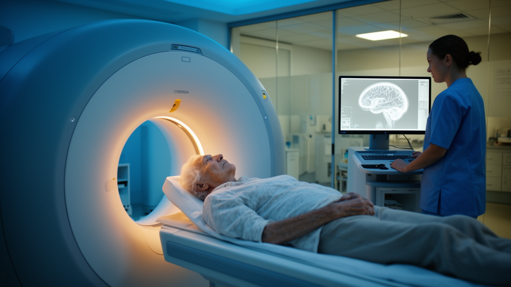
Metformin, the world’s most prescribed antidiabetic drug, appears to leave a measurable fingerprint on brain scans, according to a new translational study published in Frontiers in Radiology. Researchers at Hospital Clínico San Carlos and Universidad Complutense de Madrid in Spain found that the medication alters the distribution of 18F-fluorodeoxyglucose (18F-FDG), the radioactive glucose analog used in positron emission tomography (PET), in both mice and patients. Because 18F-FDG PET is a routine diagnostic tool for dementia-related disorders, the findings suggest that metformin treatment could potentially confound the interpretation of brain scans in the very population most likely to undergo them.
The clinical stakes are considerable. Dementia and type 2 diabetes are highly prevalent disorders that frequently coexist in elderly people, meaning a large proportion of patients referred for dementia work-ups are already taking metformin. Dementias are typically associated with characteristic patterns of brain hypometabolism: Alzheimer’s disease, the most frequent form, commonly manifests with reduced uptake in the posterior cingulate gyrus, precuneus, and posterior temporal and parietal lobes, while frontotemporal dementia is characterized by hypometabolism in the frontal and anterior temporal lobes and anterior cingulate gyrus. Any drug that shifts regional glucose metabolism could therefore mask, mimic, or distort these diagnostic signatures.
The rationale for suspecting metformin is rooted in its pharmacology. The drug lowers blood glucose primarily by inhibiting hepatic glucose production and augmenting insulin sensitivity, but it also stimulates the translocation of glucose transporter 4 (GLUT4) in an AMPK-dependent fashion. As a result, metformin can affect tissue glucose uptake, and consequently, 18F-FDG uptake. This is already well documented outside the brain: numerous PET imaging studies in metformin-treated diabetic patients and mice have shown substantially elevated tracer uptake in the bowel, which crucially impairs abdominal cancer staging by masking malignant lesions or mimicking pathologic uptake. Given that metformin readily crosses the blood-brain barrier, the Madrid team reasoned that the drug could alter cerebral 18F-FDG uptake in a similar fashion.
Previous studies on this question, however, had delivered inconclusive and even contradictory results. A retrospective clinical study in patients with type 2 diabetes reported hypermetabolism in the white matter of the temporal, frontal, and occipital lobes, alongside hypometabolism in the parahippocampal gyrus, fusiform gyrus, and ventromedial prefrontal cortex in metformin-treated individuals, which the authors attributed to neuroinflammation and glial activation. Yet more recent evidence indicates that metformin actually reduces neuroinflammation, undermining that interpretation. In contrast, a preclinical PET study in mice showed globally reduced tracer uptake after chronic metformin treatment. To resolve the uncertainty, the Spanish researchers interrogated the question in both mice and patients using complementary experimental designs.
In the preclinical arm, adult male C57BL/6J mice underwent in vivo 18F-FDG PET imaging under two treatment paradigms. In the acute paradigm, each animal served as its own control: mice received a single intraperitoneal injection of saline before a baseline scan, and then metformin at 300 mg/kg ten minutes before a second scan one week later. In the chronic paradigm, mice received metformin dissolved in drinking water at 300 mg/kg/day for three weeks, with water consumption monitored three times per week to adjust concentration, while control animals received plain water. Blood glucose levels measured before tracer injection did not differ between conditions or groups, ruling out glycemia as a confounder. Tracer uptake occurred while the animals were awake, followed by static 20-minute PET scans under isoflurane anesthesia in a dedicated small-animal PET/CT scanner.
The results were striking and region-specific. Acute metformin treatment induced significant hypermetabolism in several brain regions, including the hippocampus, where the uptake ratio rose from 0.94 ± 0.08 at baseline to 1.07 ± 0.07 after treatment (p = 0.0002), and the thalamus, which climbed from 1.00 ± 0.08 to 1.17 ± 0.08 (p = 0.0002). Significant increases were also observed in the midbrain and striatum, and statistical parametric mapping confirmed clusters of elevated voxels in hippocampus, thalamus, and subregions of cortex and striatum. Chronic treatment, by contrast, produced a more modest increase confined to cortical regions, with SPM revealing clusters mainly in rostral areas corresponding to motor and somatosensory cortex. The authors suggest that the acute effect may stem from metformin’s well-established inhibition of mitochondrial complex I, which shifts cellular energy metabolism toward glycolysis and increases glucose uptake, while the brain may adapt to sustained exposure over longer periods.
Adding a mechanistic layer, the team performed in vitro autoradiography for the translocator protein (TSPO), a marker often associated with glial activation and mitochondrial alterations, using the radioligand 3H-PK11195. Although the increase in cortical TSPO expression in chronically treated mice did not reach statistical significance, linear regression revealed a positive correlation between cortical TSPO expression and 18F-FDG uptake in a frontal cortical cluster, confirmed by Pearson’s correlation (r = 0.73, p = 0.017). The researchers speculate that this link may reflect altered mitochondrial function rather than inflammation, noting that chronic metformin treatment has recently been shown to increase mitochondrial area in neuronal axons and glial cytoplasm and to upregulate mitochondrial metabolism transcripts.
The clinical arm of the study took a retrospective approach, analyzing 18F-FDG PET images from 39 patients with non-pathological brain scans, divided into 27 untreated controls and 12 metformin-treated patients with type 2 diabetes, seven of whom were taking additional antidiabetic medication. The groups were matched in age and sex distribution, though the metformin patients had elevated glycosylated hemoglobin (HbA1c) as expected. After normalizing brain uptake to whole-brain activity and comparing the groups with SPM12, the researchers found that treated patients showed hypermetabolism in the temporal lobe, with hippocampus and parahippocampal gyrus uptake rising from 0.97 ± 0.04 to 1.03 ± 0.04 (p < 0.0001), and elevated uptake in the amygdala and white matter regions. Conversely, uptake was reduced in frontal cortical regions, including the superior frontal gyrus (1.24 ± 0.04 vs 1.28 ± 0.06; p = 0.0165), middle frontal gyrus, and inferior frontal gyrus.
Intriguingly, linear regression against HbA1c values revealed a negative correlation between the diabetes marker and 18F-FDG uptake in frontal and parietal cortical subregions, but no correlation with the hypermetabolic temporal areas. This dissociation led the authors to a nuanced interpretation: the reduced frontal uptake in their patients might be attributable to the diabetic condition itself, which has been repeatedly associated with cerebral hypometabolism in prior twin studies and comparative imaging work, whereas the hypermetabolism in white matter and temporal regions is more plausibly an effect of metformin treatment. The hippocampal findings are of particular diagnostic interest, since hypometabolism in medial temporal areas is a recognized hallmark of Alzheimer’s disease, and metformin-induced hypermetabolism in this region could potentially mask pathological patterns and complicate the differential diagnosis among neurodegenerative diseases.
The authors are careful to acknowledge the limitations of their exploratory study, including the small sample sizes, the exclusively male mouse cohorts, the supraphysiological preclinical dose, and the absence of family-wise error correction in the SPM analyses. They also note that their control group consisted of healthy individuals rather than diabetic patients without metformin, and that discrepancies with earlier studies may reflect differences in mouse strain, metformin dose, and anesthesia protocols during tracer uptake. Nevertheless, the convergence of preclinical and clinical evidence on regional alterations in brain 18F-FDG uptake carries a clear practical message: nuclear medicine clinicians should be aware of these potential metformin-related changes when evaluating brain PET scans, and new studies modifying the treatment regimen in diabetic patients prior to 18F-FDG PET imaging are warranted.
Subject of Research: Effects of metformin on cerebral 18F-FDG PET uptake in mice and diabetic patients
Article Title: Effect of metformin treatment on brain 18F-FDG uptake: a translational study
Article References: Effect of metformin treatment on brain 18F-FDG uptake: a translational study. (n.d.). https://doi.org/10.3389/fradi.2026.1917175
Image Credits: AI Generated
DOI: 10.3389/fradi.2026.1917175
Keywords: metformin, 18F-FDG PET, brain metabolism, dementia, Alzheimer's disease, type 2 diabetes, hippocampus, neuroimaging, nuclear medicine, translational research, HbA1c, mitochondrial complex I
Cite Scienmag News
Cassandra Pierce. (October 16, 2026). Metformin Reshapes Brain PET Scans, Raising New Questions for Dementia Diagnosis. Scienmag. https://scienmag.com/metformin-reshapes-brain-pet-scans-raising-new-questions-for-dementia-diagnosis/
Cassandra Pierce. "Metformin Reshapes Brain PET Scans, Raising New Questions for Dementia Diagnosis." Scienmag, 16 October 2026, https://scienmag.com/metformin-reshapes-brain-pet-scans-raising-new-questions-for-dementia-diagnosis/. Accessed 16 October 2026.
Cassandra Pierce. "Metformin Reshapes Brain PET Scans, Raising New Questions for Dementia Diagnosis." Scienmag. October 16, 2026. https://scienmag.com/metformin-reshapes-brain-pet-scans-raising-new-questions-for-dementia-diagnosis/


