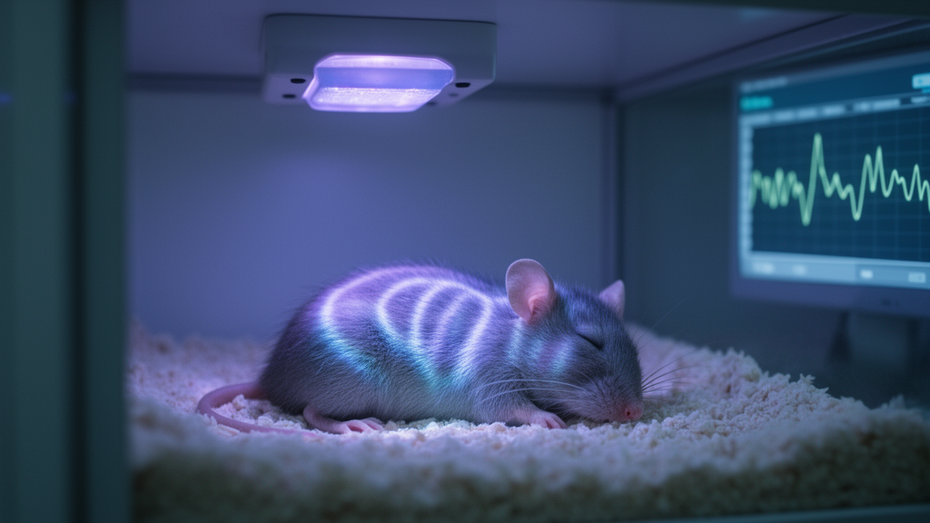
In aging brains, the timing of light therapy may matter as much as the light itself. A new study in mice reports that transcranial photobiomodulation — the delivery of near-infrared light through the skull to stimulate brain tissue — improved the clearance of beta-amyloid and boosted memory performance in old animals only when the light was applied during sleep. The same daily light course delivered during wakefulness had no measurable effect on either the molecular burden in the brain or the animals’ learning, a striking dissociation that the authors interpret as evidence that sleep opens a therapeutic window for photostimulation of brain drainage.
The work, published as an open-access research article in Frontiers of Optoelectronics, was carried out by a team at Saratov State University and collaborating institutions in Russia. It builds on their earlier finding that age is a limiting factor for photobiomodulation: while the technique reliably stimulates meningeal lymphatic vessels — the vessels that drain fluid and waste from the brain — in young animals, its effects fade in old ones. Clinical studies reviewed by the authors point in the same direction, with trials in Alzheimer’s and Parkinson’s patients over roughly 70 years of age reporting little or no cognitive or motor benefit from photobiomodulation.
To test whether sleep could restore that lost efficacy, the researchers worked with male C57BL/6 mice at three ages: 3 months, 12 months, and 24 months, which the authors map to roughly 25, 42.5, and 69 human years. The light source was a 1050-nanometer LED mounted on the skull, driven in a pulsed mode that delivered a 10 joules per square centimeter dose over 17 minutes while keeping the brain surface temperature unchanged, at 37.11 versus 37.12 degrees Celsius before and during stimulation. Because the skull thins and changes with age, the team also measured how much light actually passed through it: transmission at 1050 nanometers was 60 plus or minus 5 percent in 3-month-old mice, 47 plus or minus 5 percent at 12 months, and 39 plus or minus 5 percent at 24 months, meaning old brains received less light for the same external dose.
The central experiment compared a 10-day course of photobiomodulation delivered during EEG-verified NREM sleep against the identical course delivered during wakefulness in 24-month-old mice. Only the sleep-timed treatment changed the biology. Beta-amyloid levels in the brain fell by 1.6 times, and in the meninges by 1.4 times, relative to untreated controls, with the treated animals showing 4.80 versus 7.62 arbitrary fluorescence units in the brain and 3.83 versus 5.44 units in the meninges. Wake-timed stimulation left both measures statistically unchanged. Sham animals wearing the LED platform without active light matched the controls on every measure, ruling out effects of the hardware itself.
Cognition followed the same pattern. Using a modified Pavlovian conditioning task in which mice learn to associate a green light with a food reward, the researchers counted how many daily sessions each animal needed to form a stable reflex and how many rewards it collected. Old mice that received photobiomodulation during sleep needed 22.03 sessions on average versus 26.38 for controls, and collected 11.63 rewards versus 6.88. The wake-timed group was indistinguishable from controls on both counts. The authors chose this home-cage conditioning test deliberately, arguing in their methods discussion that conventional behavioral batteries such as the open field, novel object recognition, and Y-maze are not sensitive enough to detect age-related cognitive change — a point consistent with several prior photobiomodulation studies in aged rodents that found no effects on those tests.
A second line of experiments probed why sleep matters, using sleep deprivation as a stress test for the brain’s waste-clearance system. The team modeled acute sleep loss with a single day of gentle disturbance — introducing new objects into the cage — and chronic sleep deprivation with 3 hours of sleep exclusion per day for 10 days, a method chosen because it does not raise blood corticosterone, which the authors confirmed by ELISA in all age groups. Chronic, but not acute, sleep deprivation suppressed brain drainage in an age-graded fashion: tracer movement through the brain fell by roughly 1.7-fold in 3-month-old mice, 2- to 3.5-fold in 12-month-old mice, and up to 7.8-fold in the ventral brain of 24-month-old mice, with the deepest decline in the oldest animals whose baseline drainage was already reduced by about half compared with the young groups.
Alongside the drainage measurements, the team quantified five metabolites in brain lysates: soluble beta-amyloid 1-42, tau protein, glutamate, lactate, and glucose. Chronic sleep deprivation raised all five in every age group, but the accumulation was most pronounced in the old animals — brain beta-amyloid climbed from 11.65 to 18.30 picograms per gram of protein, tau from 92.40 to 123.35 picograms per gram, and glutamate from 44.90 to 96.11 millimoles per kilogram. Here the age divide in treatment response became stark. In young and middle-aged sleep-deprived mice, both sleep-timed and wake-timed photobiomodulation courses restored metabolite levels to near baseline. In the old mice, only the sleep-timed course worked; wake-timed stimulation failed to bring tau, glutamate, lactate, or glucose back down, leaving them significantly elevated relative to controls.
To watch the drainage process directly, the researchers implanted chronic cranial windows and intraventricular catheters in 24-month-old mice trained over one to two months to sleep naturally under a two-photon microscope without anesthesia, which is itself known to alter brain drainage. Injecting a fluorescent dextran tracer into the lateral ventricle, they tracked its movement through perivascular spaces in real time on the same animal across three days — a baseline day, a day with sleep-timed stimulation, and a day with wake-timed stimulation. Tracer occupancy in the perivascular spaces rose significantly only after sleep-timed photobiomodulation, reaching 1.58 versus 1.22 arbitrary units without stimulation, while the wake condition matched baseline. Ex vivo confocal analysis of the deep cervical lymph nodes, the first collection station for fluid leaving the brain, showed the same selectivity: tracer accumulated there only in the sleep-stimulated group.
The authors are careful about mechanism. They note that whether the glymphatic system — the proposed convective flow of fluid through perivascular spaces driven by arterial pulsation and aquaporin channels — exists as described remains actively debated, and that direct lymphatic vessels within the brain parenchyma have likewise not been conclusively demonstrated. What their data show is that sleep-timed light increases both fluid movement in perivascular spaces and tracer delivery to cervical lymph nodes in old mice, consistent with the idea that the natural activation of brain drainage during sleep provides the flowing substrate that photostimulation needs in order to push clearance. In young brains, they suggest, meningeal lymphatic function is preserved enough that wake-timed light still works; in old brains, where the vessels show hyperplasia and valve dysfunction, only the sleep state supplies sufficient baseline flow for the light to amplify.
The study’s limitations are those of the model. All findings come from male mice, with eight animals per group in the main experiments and five in the imaging cohorts, and the translation from a 24-month-old mouse to a human patient over 70 is an extrapolation the authors themselves flag as approximate. They also acknowledge that they deliberately kept the light dose constant across ages despite the age-related drop in skull transmission, and that simply raising the dose may not compensate, based on their earlier work showing that dose is not the decisive variable for drainage effects. What the results establish is narrower but concrete: in aged mice, the efficacy of transcranial photobiomodulation for clearing beta-amyloid, restoring sleep-deprivation-induced metabolite accumulation, and improving conditioned learning depends on delivering the light during sleep. If the sleep-timed approach transfers to clinical settings, it would suggest that the long-sought way to make light therapy work in the elderly brain may lie not in stronger light, but in better timing.
Subject of Research: Photobiomodulation during sleep enhances brain waste clearance and cognition in aged mice
Article Title: Sleep is a therapeutic window for photostimulation of drainage of aging brain
Article References: Andrey, T., Viktoria, A., Alexander, S., Nikita, N., Inna, B., Daria, Z., Anastasiia, S.-G., Sonina, K., Arina, E., Inna, E., Matvey, T., Alexander, D., Alexander, D., Dmitry, M., Sergey, P., Dmitry, T., Egor, I., Maria, T., Ivan, F., & Oxana, S.-G. (2025). Sleep is a therapeutic window for photostimulation of drainage of aging brain. Frontiers of Optoelectronics, 18(1), Article 22. https://doi.org/10.1007/s12200-025-00168-0
Image Credits: AI Generated
DOI: 10.1007/s12200-025-00168-0
Keywords: photobiomodulation, sleep, aging brain, meningeal lymphatic vessels, beta-amyloid, glymphatic system, sleep deprivation, brain drainage, cognitive function, near-infrared light, neurodegeneration, mouse study
Cite Scienmag News
Beatrice Stafford. (October 28, 2026). Light Therapy Works Only During Sleep in Aging Mouse Brains. Scienmag. https://scienmag.com/light-therapy-works-only-during-sleep-in-aging-mouse-brains/
Beatrice Stafford. "Light Therapy Works Only During Sleep in Aging Mouse Brains." Scienmag, 28 October 2026, https://scienmag.com/light-therapy-works-only-during-sleep-in-aging-mouse-brains/. Accessed 28 October 2026.
Beatrice Stafford. "Light Therapy Works Only During Sleep in Aging Mouse Brains." Scienmag. October 28, 2026. https://scienmag.com/light-therapy-works-only-during-sleep-in-aging-mouse-brains/