

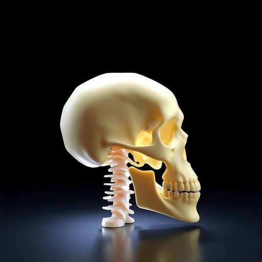
For infants born with craniosynostosis, the skull’s soft, growing seams fuse into rigid bone far too early, trapping the brain inside a closing vault. The condition affects roughly one in every 2,500 births and can trigger a cascade of problems: distorted head shape, elevated intracranial pressure, developmental delays, and the prospect of multiple high-risk surgeries throughout childhood. Even after surgeons carve open the fused sutures, the bone often knits itself back together, demanding further intervention. Now, a team of bioengineers and developmental biologists has taken a radically different approach. Instead of merely cutting bone away, they have built a biodegradable scaffold that regenerates the very stem cell niche the skull needs to keep growing — and in mice, it stopped re-fusion while restoring craniofacial development.
The work, published in Bone Research, was led by Professor Yuji Mishina at the University of Michigan School of Dentistry and Dr. W. Benton Swanson at the Harvard School of Dental Medicine. Their central insight was that craniosynostosis is not just a bone overgrowth problem; it is a stem cell extinction event. Cranial sutures normally house a reservoir of skeletal stem cells that orchestrate the lifelong balance between bone deposition and fibrous tissue maintenance. When disease signals obliterate that niche, the suture collapses into a solid bony bridge. “Our goal was not simply to reopen a fused suture, but to regenerate the biological niche that allows the skull to grow normally,” Mishina explained. To do that, the researchers designed a triphasic scaffold that mimics the natural “bone-suture-bone” architecture.
The implant is fabricated entirely from poly(L-lactic acid), an FDA-approved polymer already used in dissolvable medical devices, but its internal geometry is what makes it a niche-regenerating machine. The scaffold contains three distinct compartments with precisely tuned pore sizes. A central band of small pores creates a protective microenvironment that preserves skeletal stem cells in an undifferentiated state. On either side, larger pores encourage blood vessel infiltration and osteogenic differentiation, giving rise to new bone. This spatial segregation directly channels cell fate: stem cells placed in the middle zone retained their stemness markers and self-renewal capacity, while their progeny that received differentiation cues migrated outward and contributed to mineralized tissue. Lineage-tracing confirmed that the scaffold maintained a functional stem cell reservoir while allowing descendants to participate in regeneration — a dynamic that closely mirrors healthy suture biology.
The team then stress-tested the system against the biological drivers of craniosynostosis. They exposed the scaffolds to excessive bone morphogenetic protein (BMP) signaling, a pathway notorious for triggering pathological bone formation. In conventional settings, such signaling would rapidly ossify any cell-laden construct. Yet the central small-pore compartment resisted calcification and preserved a non-bony, stem-cell-rich niche, demonstrating that the physical microenvironment can override potent pro-osteogenic cues. Even when bathed in the molecular signals that normally seal sutures shut, the engineered niche maintained an open, soft-tissue identity.
Moving into a mouse model of midline craniosynostosis — the most common nonsyndromic form in humans — the researchers surgically removed the fused suture and implanted the triphasic scaffold. Animals that received a standard surgical repair, without the niche-regenerating implant, experienced the familiar re-fusion. In contrast, those with the scaffold retained a patent, suture-like tissue that did not ossify. Detailed microcomputed tomography and histology revealed improved skull dimensions and more normal craniofacial growth trajectories. Early intervention yielded the most pronounced benefits, underscoring the importance of restoring stem cell function during critical developmental windows.
Swanson emphasized the broader design logic behind the achievement. “This work demonstrates how rational biomaterial design can control stem cell fate and tissue organization simultaneously,” he said. “We believe the principles established here may be broadly applicable to regenerative therapies beyond craniosynostosis.” Indeed, the concept of building physical compartments that parse self-renewal from differentiation could inform next-generation implants for long bone defects, cartilage repair, or even soft organ regeneration where stem cell maintenance is crucial.
The study also offers a glimpse of a future where complex congenital defects are treated not with static patches but with living, responsive niches that guide tissue growth. While human trials remain on the horizon, the poly(L-lactic acid) scaffold’s existing regulatory track record may smooth the translational path. For families facing the relentless cycle of craniosynostosis surgeries, a single bioengineered implant that coaxes the skull to keep growing properly represents a profound shift — one that moves from cutting away bone to rebuilding the body’s own developmental machinery.
Subject of Research: Animals
Article Title: A tissue engineering approach to regenerate the cranial suture skeletal stem cell niche with a multicompartment biomaterial scaffold
News Publication Date: 28-May-2026
Web References: http://dx.doi.org/10.1038/s41413-026-00539-z
References: Bone Research, 2026; 14: DOI: 10.1038/s41413-026-00539-z
Image Credits: Dr. W. Benton Swanson from Harvard University, USA, and Professor Yuji Mishina from the University of Michigan, USA
Keywords: craniosynostosis, skeletal stem cells, tissue engineering, biomaterial scaffold, bone morphogenetic protein, skull regeneration, stem cell niche, cranial sutures, poly(L-lactic acid), craniofacial development


