


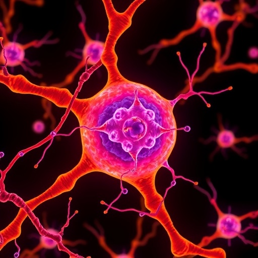
Microglia, the sentinel immune cells inhabiting the brain, are essential guardians of neural health. Their dynamic ability to remodel the internal framework known as the actin cytoskeleton plays a crucial role in maintaining brain homeostasis by facilitating the removal of unwanted substances. Central to this remodeling capacity is a proton channel protein named Hv1/VSOP, widely recognized for its expression on microglial cell membranes where it modulates extracellular pH through proton transport. However, groundbreaking research has unveiled an unprecedented role for Hv1, extending beyond its traditional localization to the cell surface and uncovering its functional presence on intracellular transport vesicles known as endosomes.
The discovery that Hv1 operates on endosomal membranes was made possible through the application of cutting-edge microscopy paired with an innovative technique called endosome patch-clamping. This method enables direct measurement of ionic currents through microscopic membrane structures deep within cells, a feat previously unattainable. Combining this approach with advanced optical imaging, researchers demonstrated that Hv1 is not a mere cell surface channel but an active proton conduit on endosomes, fundamentally altering our understanding of its intracellular dynamics and functions.
The implications of Hv1’s presence on endosomes are profound. Microglia deficient in Hv1 exhibit significant cellular abnormalities, particularly in the morphology and function of their actin cytoskeleton. Without Hv1, the actin filaments elongate excessively, leading to distorted cell shapes and impaired internal organization. This aberrant growth suggests that Hv1 acts as a molecular brake, suppressing unchecked actin polymerization and preserving cellular architecture essential for microglial function.
Delving deeper into the molecular machinery, detailed biochemical analyses revealed direct interactions between Hv1 and a protein called CAPZ. CAPZ is known for its role in binding to the barbed ends of actin filaments, effectively capping them to limit filament elongation. The partnership between Hv1 and CAPZ indicates a novel regulatory axis where ion channel activity on endosomes influences the dynamic remodeling of the cytoskeleton. This interaction modulates actin filament growth precisely, ensuring that cellular shape and motility remain finely tuned.
Live-cell imaging provided compelling visual confirmation of this mechanism. Endosomes adorned with Hv1 were observed physically associating with the tips of actin filaments. This spatial coupling points to a scenario where endosomal Hv1 guides cytoskeletal dynamics from within, orchestrating the organization and turnover of actin structures in real time. It challenges the prevailing paradigm that ion channels predominantly function at the plasma membrane, positioning Hv1 as a key intracellular regulator.
Such findings illuminate a sophisticated cellular control system whereby ion channels on endosomal membranes are integral to the formation and spatial arrangement of the actin cytoskeleton. This convergence of ion transport and cytoskeletal regulation underscores the intricate cross-talk between intracellular signaling and structural adaptation, vital processes underpinning microglial surveillance and response.
Moreover, Hv1’s role on endosomes extends beyond mere structural influence; it implicates proton flux as a signaling modality within intracellular compartments. By modulating local pH near actin filament ends, Hv1 may affect CAPZ’s ability to cap filaments and thereby finely adjust actin dynamics in response to cellular states. This adds an additional layer of complexity to how microglia adapt their morphology and function during health and disease.
The broader implications of this research ripple across neurobiology and immunology. Microglia’s capability to maintain brain health hinges on their shape-shifting abilities, driven by cytoskeletal remodeling. Understanding Hv1’s unexpected intracellular role opens potential avenues for targeted therapies aimed at modulating microglial behavior in neurodegenerative diseases, where aberrant immune activation and cytoskeletal dysfunction contribute to pathology.
Technically, this study bridges specialized electrophysiology with cell biology, leveraging the precision of patch-clamp recordings at the nanoscale level of endosomes. Such integration paves the way for future inquiries into intracellular ion channel functions, revealing layers of cellular regulation that were previously obscured by technical limitations.
In summary, the identification of Hv1’s active proton channel functionality on endosomes and its regulatory influence over the actin cytoskeleton via CAPZ interaction represents a paradigm shift. It transforms our understanding of microglial biology and unveils an unexplored nexus of ion channel signaling and cytoskeletal organization, with far-reaching implications for brain health and immunity.
This remarkable body of work not only expands our comprehension of cellular physiology but also sets a new foundation for exploring how intracellular ion channels contribute to cell morphology, signaling pathways, and immune cell functions. It highlights the importance of a holistic view of ion channel distribution and function—beyond membranes facing the extracellular environment—to unlock the full spectrum of cellular complexity.
As research moves forward, the challenge will be to map the precise molecular mechanisms linking Hv1 activity, proton flux, and CAPZ regulation in varying physiological and pathological contexts. Unraveling these connections may illuminate novel targets for modulating microglial activity in diverse neurological disorders, underscoring the translational potential of this fundamental discovery.
Cite Scienmag News
Drew Townsend. (May 14, 2026). Ion Channels Within Cells Actively Remodel the Cytoskeleton. Scienmag. https://scienmag.com/ion-channels-within-cells-actively-remodel-the-cytoskeleton/
Drew Townsend. "Ion Channels Within Cells Actively Remodel the Cytoskeleton." Scienmag, 14 May 2026, https://scienmag.com/ion-channels-within-cells-actively-remodel-the-cytoskeleton/. Accessed 6 September 2026.
Drew Townsend. "Ion Channels Within Cells Actively Remodel the Cytoskeleton." Scienmag. May 14, 2026. https://scienmag.com/ion-channels-within-cells-actively-remodel-the-cytoskeleton/


