In a groundbreaking study from the University of Tokyo, researchers have unveiled critical insights into how the membrane lipid composition of extracellular vesicles (EVs) governs their surface charge. This discovery sheds light on the fundamental differences between subtypes of EVs, notably exosomes and membrane-derived vesicles, attributing these distinctions primarily to the asymmetric distribution of specific phospholipids such as phosphatidylserine. This pivotal research not only deepens our understanding of EV biology but also has profound implications for the development and standardization of EV-based diagnostic tools and therapeutic agents.
Extracellular vesicles are small, membrane-bound particles secreted by cells, playing vital roles in intercellular communication by ferrying proteins, nucleic acids, and lipids. They have attracted considerable attention in recent years due to their potential in disease diagnostics, drug delivery, and as therapeutic agents. However, the heterogeneity among EV subpopulations—in terms of size, content, and biogenesis pathways—has complicated their classification and functional understanding. The work led by the Innovation Center of NanoMedicine (iCONM) as part of the JST COI-NEXT program addresses one of the central challenges: identifying reliable markers that distinguish EV subtypes based on fundamental physicochemical properties.
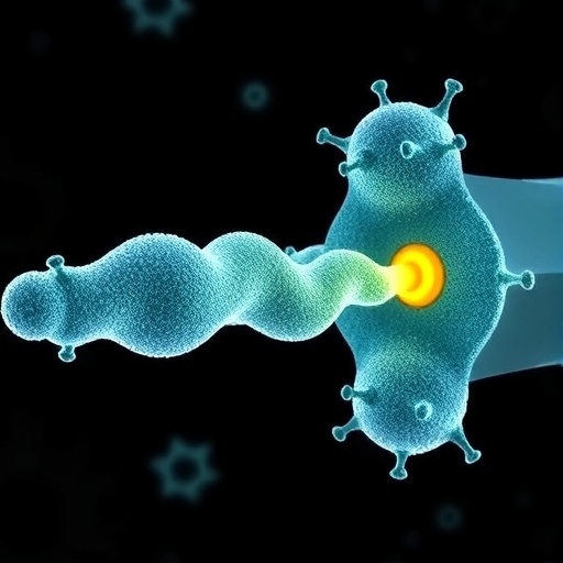
At the heart of this research lies the concept of phospholipid asymmetry in the vesicle membrane. The asymmetric distribution refers to the uneven localization of various phospholipids between the inner and outer leaflets of the lipid bilayer. Phosphatidylserine, one such negatively charged phospholipid, is typically confined to the inner leaflet in healthy cells but can be externalized during EV formation. The researchers demonstrated that this asymmetric distribution critically influences the vesicle’s surface potential, otherwise known as the zeta potential. By comparing the lipid composition and zeta potentials of exosomes and membrane-derived EVs, they provided compelling evidence linking phosphatidylserine exposure to differences in electric charge on the vesicle surface.
The significance of zeta potential in this context cannot be overstated. Zeta potential is a measure of the electric potential at the slipping plane of a particle suspended in fluid and is widely used to infer surface charge properties. For extracellular vesicles, surface charge heavily dictates their interaction with the extracellular environment, cellular uptake efficiency, and biodistribution in vivo. This study advocates for adopting zeta potential as a practical biophysical parameter for EV classification, one which could streamline quality control processes and ensure consistency in EV production—a major bottleneck in their translational application.
Methodologically, the team employed advanced lipidomic analyses combined with electrophoretic mobility measurements to quantify and compare the phospholipid profiles and zeta potentials of isolated EV populations. This integrative approach allowed for the precise delineation of membrane composition differences underpinning the variations in surface charge. Such technical rigor not only reinforces the validity of their findings but also sets a new standard for experimental protocols in EV research, encouraging more nuanced investigations that consider the intricate lipid landscape of vesicles.
Beyond diagnostics and classification, the findings harbor critical implications for the design of EV-based therapeutics. Therapeutic EVs must possess predictable and reproducible physicochemical characteristics to be safe and efficacious. Understanding how membrane lipid asymmetry modulates surface charge provides a tangible parameter to rationally engineer EVs with customized interactions for target cell uptake or circulation stability. This capability could revolutionize the field of nanomedicine by enabling the fine-tuning of vesicle surface properties to optimize therapeutic payload delivery and minimize off-target effects.
Furthermore, the research paves the way for standardized protocols which are indispensable for regulatory approval of EV therapies. Currently, the lack of universally accepted criteria for EV characterization hinders their commercialization and clinical translation. By proposing zeta potential as a quantitative marker aligned with membrane lipid asymmetry, this work offers a viable foundation for regulatory guidelines that ensure batch-to-batch uniformity and quality. This standardization could accelerate EVs’ journey from laboratory curiosities to mainstream therapeutics.
The insights uncovered also extend to understanding the physiological roles of EV subtypes within the body. Since surface charge influences vesicle interactions with immune cells, extracellular matrix components, and plasma proteins, differential zeta potentials among EV categories likely reflect functional adaptations. Exosomes, characterized by distinct phosphatidylserine distributions and negative surface charge, might exhibit unique biodistribution profiles or immune evasion strategies compared to other EV forms. Consequently, this study reframes how researchers conceptualize EV heterogeneity from a biophysical perspective linked intimately to membrane chemistry.
This exciting breakthrough was achieved within the Innovation Center of NanoMedicine (iCONM), a collaborative hub aiming to propel nanotechnological advances in medicine. The JST COI-NEXT program’s support highlights the strategic importance of interdisciplinary approaches uniting lipidomics, nanotechnology, and clinical science. By fostering such collaborative environments, the University of Tokyo team has demonstrated how crossing traditional boundaries can yield transformative knowledge that reshapes biomedical paradigms.
Looking forward, the research community anticipates that these findings will catalyze a wave of studies exploring how manipulating lipid asymmetry can tune EV functionality. Potential future directions include the development of synthetic vesicles mimicking natural phospholipid distributions to achieve targeted delivery or the creation of diagnostic platforms leveraging zeta potential measurements for rapid EV subtype screening. Ultimately, the elucidation of charge determinants at the nanoscale will enrich both fundamental cell biology and applied therapeutic design.
In sum, this seminal work offers a clarifying lens on the molecular underpinnings that differentiate extracellular vesicle subclasses. By pinpointing the role of phosphatidylserine-driven lipid asymmetry in defining surface charge, the University of Tokyo researchers have provided a robust, quantifiable target for EV classification and quality control. This advance promises to accelerate the realization of EVs as reliable, standardized tools in the burgeoning fields of diagnostics and nanomedicine, marking a transformative moment in vesicle research.
Subject of Research: Extracellular vesicle membrane lipid composition and surface charge characterization
Article Title: (Not provided)
News Publication Date: (Not provided)
Web References: (Not provided)
References: (Not provided)
Image Credits: University of Tokyo / Innovation Center of NanoMedicine (iCONM)
Keywords: extracellular vesicles, membrane lipid asymmetry, phosphatidylserine, zeta potential, exosomes, nanomedicine, EV classification, quality control, lipidomics, therapeutic design, JST COI-NEXT
