


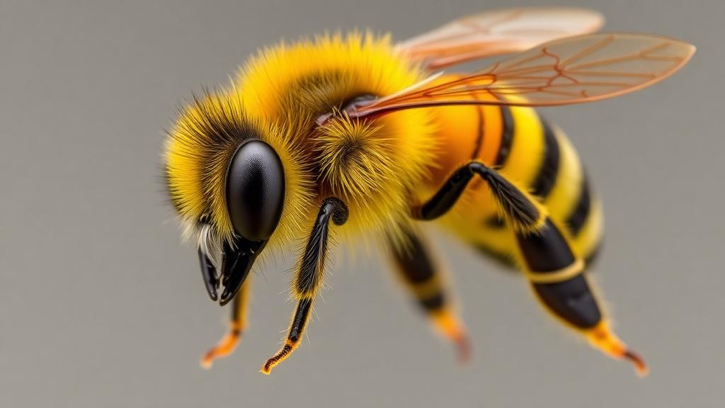
The honey bee sting is one of the most familiar minor injuries in the natural world, and yet the mechanism behind it has remained surprisingly mysterious for more than a century. Scientists have known since the early twentieth century that venom flows from a sac into a chamber equipped with small pumping elements, but the precise way those elements generate pressure has never been fully explained. Now a team of Australian researchers has captured the process in extraordinary detail, showing that the bee’s stinger is not a passive needle but a self-contained, self-powered micropump that keeps working even after it has been torn from the bee’s own body.
The new study, published in the Proceedings of the National Academy of Sciences, was led by Dr Fiorella Ramirez-Esquivel of Macquarie University, working with colleagues at Macquarie and the University of New South Wales. Using high-speed videography, the team filmed the stinger of the honey bee Apis mellifera as it pumped venom into a transparent gel designed to mimic human skin. The recordings, captured at 300 frames per second, revealed for the first time the complete pumping cycle of a structure barely two millimetres long, exposing a level of mechanical sophistication that the researchers describe as remarkable for such a tiny organ.
The story begins with a behavior most people have witnessed at least secondhand. When a worker honey bee stings a target with sufficiently elastic skin, she thrusts the stinger in and then flies away, leaving the entire stinging apparatus behind. This process, known as autotomy, is fatal to the bee, but it serves the colony by ensuring that venom delivery continues long after the bee herself is gone. The detached stinger does not simply sit in the wound. It continues to work its way deeper into the tissue and keeps injecting venom under its own power, driven by the muscles, nerves and mechanical components packed into the tiny structure.
To observe this autonomous action, the researchers had bees sting a clear gel that mimics the mechanical properties of human skin. Because the substrate is transparent, transmitted-light illumination allowed the team to film the internal workings of the stinger as it operated. The footage shows the autotomised stinger advancing through the phantom tissue through the reciprocating action of its two lancets, the paired, sharply pointed elements that form the piercing tip. In the artificial substrate, which cannot absorb or wick away venom, the injected fluid is eventually forced back up to the surface, making the pumping action visible in striking detail.
The stinger’s anatomy is a compact assembly of cooperating parts. It contains a venom sac that empties into a bulb-shaped pumping chamber, along with the muscles and nerves that drive the mechanism. Two long lancets slide backwards and forwards along a system of rails called the olistheter, a motion the researchers compare to that of a reciprocating saw. These lancets perform two jobs at once: they alternately drive the stinger deeper into the skin, and they pump venom from the chamber down into the wound. Crucially, each lancet carries a valve that sits inside the pumping chamber, and it is these valves that turn out to be the heart of the system.
High-speed video showed that the valves are not rigid flaps but dynamic, shape-changing structures. On the downstroke, each valve inflates like a tiny parachute, expanding to form a seal against the wall of the chamber and functioning as a piston that applies downward pressure on the venom. On the upstroke, the valve collapses, allowing venom to flow into the chamber from the sac while minimizing backflow. This alternating inflation and collapse means each valve behaves as a collapsible piston, sealing when it needs to push and opening when it needs to refill, a solution to the fundamental pumping problem of maintaining pressure without losing fluid on the return stroke.
Perhaps the most dramatic discovery concerns what happens at the midpoint of each stroke. As the two valves meet in the middle of the pumping chamber, they change shape again, deforming to push past one another. Dr Ramirez-Esquivel notes that maintaining a seal is vital for effectively transmitting pressure, and this ability of the valves to deform and slide past each other while remaining sealed is a remarkable strategy for moving fluid. Because the two pistons operate in opposite directions, one is always delivering pressure while the other refills, producing a continuous flow of venom rather than the pulsed output a single piston would generate.
The team did not rely on observation alone. To probe the mechanics of the system, they used micro-computed tomography scans of the stinger to build a three-dimensional picture of the bulb chamber and lancets, then 3D printed an upscaled working model of the mechanism. This small robot reproduced the pumping cycle and confirmed the key features seen in the high-speed footage, providing experimental support for the interpretation of the valve dynamics. The combination of direct video evidence and a physical model gives the study an unusually complete account of how a biological micropump actually works.
Dr Ramirez-Esquivel suggests that the double-piston design adds complexity, but that this complexity is the key to the stinger’s ability to deliver venom continuously in a compact, autonomous unit. The entire device, including its power source in the form of muscles and its control system in the form of nerves, operates without any connection to the bee after autotomy. That independence is what makes the stinger so effective as a defensive weapon, and it is also what makes the mechanism so interesting to engineers. A pump that can seal, deform, refill and deliver continuously within a two-millimetre envelope is a feat that conventional engineered micropumps struggle to match.
The authors conclude that the design principles uncovered in the honey bee stinger could inspire new engineered micropumps, with potential applications in minimally invasive medical devices and microfluidic technologies, fields where delivering small, controlled volumes of fluid through tiny openings is a central challenge. A mechanism refined by millions of years of evolution, in which collapsible pistons inflate to seal and collapse to refill while sliding past each other in a shared chamber, offers a template for pumps that are smaller, simpler and more reliable than many current designs. What has been causing pain for as long as humans have walked the earth may, thanks to 300-frames-per-second video and a 3D-printed robot bee stinger, end up improving drug delivery and diagnostic devices of the future.
Subject of Research: The biomechanical mechanism of venom pumping in the honey bee stinger
Article Title: Ouch! The stinger of a honey bee is revealed as a tiny autonomous double-piston venom pump
Article References: Ouch! The stinger of a honey bee is revealed as a tiny autonomous double-piston venom pump. (n.d.). Original publication
Image Credits: AI Generated
DOI: Not provided
Keywords: honey bee, stinger, venom, autotomy, double-piston pump, high-speed video, microCT, biomechanics, micropump, biomimetics, microfluidics, PNAS
Cite Scienmag News
Drew Townsend. (October 12, 2026). Honey Bee Stinger Revealed as Tiny Autonomous Double-Piston Venom Pump. Scienmag. https://scienmag.com/honey-bee-stinger-revealed-as-tiny-autonomous-double-piston-venom-pump/
Drew Townsend. "Honey Bee Stinger Revealed as Tiny Autonomous Double-Piston Venom Pump." Scienmag, 12 October 2026, https://scienmag.com/honey-bee-stinger-revealed-as-tiny-autonomous-double-piston-venom-pump/. Accessed 12 October 2026.
Drew Townsend. "Honey Bee Stinger Revealed as Tiny Autonomous Double-Piston Venom Pump." Scienmag. October 12, 2026. https://scienmag.com/honey-bee-stinger-revealed-as-tiny-autonomous-double-piston-venom-pump/


