


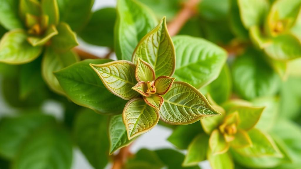
A new laboratory study has found that a methanolic extract of holy basil leaves, a plant long revered in traditional medicine, can suppress the metabolic activity of MCF-7 human breast cancer cells and trigger a cascade of changes in the cells’ redox chemistry. The research, published in BMC Complementary Medicine and Therapies by Emmanuel Udochukwu Obasi of the Federal University of Medical and Health Sciences in Kwale, Nigeria, together with Parker Elijah Joshua and Victor N. Ogugua of the University of Nigeria, Nsukka, offers a careful, preliminary portrait of how one fraction of Ocimum sanctum behaves when applied directly to cancer cells in culture. The authors are quick to stress that their findings are a starting point rather than a proof of mechanism, but the work adds to a growing body of evidence that phytochemical-rich plant preparations can perturb the delicate biochemical balance that cancer cells rely upon to survive.
The team began by dividing compounds from holy basil leaves into three solvent fractions: hexane, ethyl acetate, and methanol. Sequential extraction with solvents of increasing polarity is a standard strategy in natural products chemistry, because different classes of plant metabolites dissolve preferentially in different solvents. Non-polar compounds such as long-chain hydrocarbons tend to partition into hexane, moderately polar phenolics and terpenoids into ethyl acetate, and more polar molecules into methanol. Each fraction was then tested on MCF-7 cells, an estrogen-receptor-positive breast cancer cell line that has served as a workhorse of breast cancer research since the 1970s. Cytotoxicity was measured with the WST-8 assay, a colorimetric test in which living cells convert a tetrazolium salt into a colored formazan product; the intensity of the signal serves as a proxy for metabolic activity and, by extension, cell viability.
Of the three fractions, the methanolic extract, abbreviated OSMF, produced the strongest reduction in the WST-8 metabolic signal. The researchers estimated an apparent IC50 value, the concentration that halves the metabolic readout, of 736.7 micrograms per milliliter. That figure is comparatively high, which means the extract is only moderately potent on a per-mass basis, and the authors also observed that the dose-response relationship at the highest concentration was non-monotonic, meaning the signal did not fall in a simple, linear fashion as the dose rose. Such non-monotonic behavior is not unusual for crude plant extracts, which contain mixtures of dozens of compounds that may act antagonistically or with varying kinetics. Nevertheless, because OSMF outperformed the hexane and ethyl acetate fractions, it was selected for all subsequent experiments.
With OSMF in hand, the researchers exposed MCF-7 cells to two concentrations, corresponding to 50 percent and 100 percent of the apparent IC50, for two durations, 24 and 48 hours. They then measured a panel of redox-associated biochemical markers using standard colorimetric and enzymatic assays. The markers were chosen because cellular redox balance, the equilibrium between oxidants and antioxidants, is intimately linked to cell survival, stress adaptation, and programmed cell death. Cancer cells in particular often maintain a shifted redox state that supports their uncontrolled proliferation, making the antioxidant machinery an attractive target for investigation, even if the precise relationship between redox perturbation and cell death remains an open question in the field.
The results were striking in one direction. Rather than accumulating damage, the treated cells showed reduced levels of malondialdehyde, or MDA, a lipid peroxidation product commonly used as an indicator of oxidative damage to cell membranes. At the same time, levels of reduced glutathione, the cell’s principal small-molecule antioxidant, rose, as did the activities of four antioxidant enzymes: superoxide dismutase, catalase, glutathione S-transferase, and glutathione peroxidase. Superoxide dismutase converts the superoxide radical into hydrogen peroxide, catalase and glutathione peroxidase then break hydrogen peroxide down into water, and glutathione S-transferase conjugates reactive electrophiles to glutathione for detoxification. A coordinated increase across this entire enzymatic armory, alongside reduced lipid peroxidation, suggests the cells mounted a defensive antioxidant response to the extract rather than suffering an overwhelming oxidative assault.
Nitric oxide-associated responses told a more limited story. The researchers measured nitrite levels as an indirect readout of nitric oxide, a signaling molecule involved in inflammation, vascular regulation, and cell death pathways. Nitrite-associated signals showed only a restricted response across the treatment conditions, with the clearest increase observed at a single treatment condition rather than a consistent dose- or time-dependent pattern. The authors are explicit that these data do not establish inducible nitric oxide synthase-dependent signaling, a molecular pathway that would require direct enzymatic or gene-expression evidence to confirm. The cautious framing reflects a broader principle in cell biology: indirect colorimetric readouts can suggest, but never prove, the activity of specific molecular pathways.
Perhaps the most visually compelling evidence came from the microscope. Under light microscopy, treated MCF-7 cells displayed shrinkage, rounding, membrane blebbing, and detachment from the culture surface. These are classic morphological hallmarks associated with apoptosis, the orderly form of programmed cell death in which cells dismantle themselves without provoking inflammation. Membrane blebbing in particular, in which the cell surface forms irregular bulges as the cytoskeleton collapses, is one of the most recognizable visual signatures of the apoptotic program. However, the authors emphasize that morphology alone is not molecular confirmation. Apoptosis, necrosis, and other forms of cell death can share overlapping visual features, and distinguishing among them requires molecular markers such as caspase activation, annexin V staining, or DNA fragmentation assays, none of which were performed in this study.
To begin characterizing what might be responsible for the observed effects, the team analyzed OSMF by gas chromatography-mass spectrometry, or GC-MS, a technique that separates volatile compounds and identifies them by matching their fragmentation spectra against spectral libraries. The analysis identified several putative constituents, including 2,4-di-tert-butylphenol, a phenolic compound with reported antioxidant and bioactive properties, and 2,6,10-trimethyldodecane, a branched hydrocarbon. The word putative is doing important work here: GC-MS library matching provides tentative identifications that would need to be confirmed with authentic standards, and the technique misses non-volatile compounds entirely, which means the true chemical composition of the methanolic fraction is likely richer than the detected list suggests. Phenolic compounds in particular, many of which are poorly volatile, are known to be abundant in Ocimum sanctum and are plausible contributors to biological activity.
The authors are admirably transparent about the limits of their work, and their conclusion section reads as a roadmap for what must come next. The present findings, they write, do not establish direct reactive oxygen species generation, iNOS-dependent signaling, or molecularly confirmed apoptosis. To define the mechanism and selectivity of the observed response, they call for direct ROS measurements using fluorescent probes, molecular cell-death markers, orthogonal viability assays that do not rely solely on metabolic activity, and, critically, studies in non-tumorigenic breast cells. That last point addresses the central question of selectivity: an extract that kills cancer cells is only interesting if it spares healthy ones, and without a parallel comparison in normal mammary epithelial cells, the therapeutic relevance of the cytotoxicity remains unknown. The relatively high IC50 value also raises the practical question of whether such concentrations could ever be achieved in living tissue.
Still, the study exemplifies a disciplined approach to a field often crowded with overclaiming. Ocimum sanctum, known commonly as holy basil or tulsi, has been investigated for antioxidant, cytotoxic, and pharmacological activities for years, but the cellular responses associated with its solvent fractions have required further characterization, as the authors note in their background. By systematically comparing three fractions, quantifying a broad panel of redox markers, documenting morphological changes, and profiling constituents, the Nigerian team has generated a coherent preliminary dataset that others can build upon. The paradoxical combination of reduced lipid peroxidation with elevated antioxidant defenses in dying cells is itself intriguing, hinting that the extract may induce stress responses that ultimately fail to save the cell, or that the observed redox shifts reflect adaptation rather than damage. Untangling that puzzle will require the very experiments the authors propose. For now, the work stands as a measured contribution to redox biology and natural products research, a reminder that even beloved traditional remedies must pass through the slow, skeptical machinery of cell biology before any claim of anticancer activity can be taken seriously.
Subject of Research: Cytotoxicity and redox-associated biochemical responses to the methanolic leaf fraction of Ocimum sanctum in MCF-7 human breast cancer cells
Article Title: Cytotoxicity and redox-associated responses to the methanolic fraction of Ocimum sanctum in MCF-7 breast cancer cells
Article References: Obasi, E. U., Joshua, P. E., & Ogugua, V. N. (2026). Cytotoxicity and redox-associated responses to the methanolic fraction of Ocimum sanctum in MCF-7 breast cancer cells. BMC Complementary Medicine and Therapies. https://doi.org/10.1186/s12906-026-05620-y
Image Credits: AI Generated
DOI: 10.1186/s12906-026-05620-y
Keywords: Ocimum sanctum, holy basil, MCF-7, breast cancer, cytotoxicity, WST-8 assay, redox homeostasis, antioxidant enzymes, glutathione, malondialdehyde, apoptosis, GC-MS
Cite Scienmag News
Nathaniel Bowman. (September 30, 2026). Holy Basil Extract Shows Cytotoxic and Redox Effects in Breast Cancer Cells. Scienmag. https://scienmag.com/holy-basil-extract-shows-cytotoxic-and-redox-effects-in-breast-cancer-cells/
Nathaniel Bowman. "Holy Basil Extract Shows Cytotoxic and Redox Effects in Breast Cancer Cells." Scienmag, 30 September 2026, https://scienmag.com/holy-basil-extract-shows-cytotoxic-and-redox-effects-in-breast-cancer-cells/. Accessed 17 October 2026.
Nathaniel Bowman. "Holy Basil Extract Shows Cytotoxic and Redox Effects in Breast Cancer Cells." Scienmag. September 30, 2026. https://scienmag.com/holy-basil-extract-shows-cytotoxic-and-redox-effects-in-breast-cancer-cells/


