


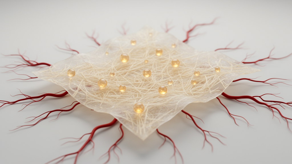
Deep within the scaffolding that holds our tissues together, researchers have found a population of tiny vesicles that may hold the key to coaxing damaged tissue into growing new blood vessels. In a study published in Frontiers in Bioengineering and Biotechnology, a team of scientists from Laval University in Canada and the University of Piemonte Orientale in Italy reports that matrix bound nanovesicles, or MBVs, extracted from decellularized bovine pericardium can drive angiogenesis in laboratory cultures and, strikingly, rescue limbs from tissue death in a mouse model of severe ischemia. The findings position these nanoscale messengers as a promising new tool for therapeutic angiogenesis, the regenerative medicine strategy that aims to restore blood supply to oxygen-starved tissue.
MBVs are a relatively recent discovery in the world of extracellular vesicles. Unlike exosomes and microvesicles, which circulate freely in body fluids, MBVs remain embedded within the collagen component of the extracellular matrix, the fibrous scaffold that surrounds and supports cells in every tissue. First identified in 2016 by researchers working with decellularized bioscaffolds, these vesicles have since been isolated from a variety of decellularized tissues and shown to carry a rich, tissue-specific cargo of proteins, lipids, and nucleic acids. Their ability to modulate immune responses and promote tissue remodeling has already been demonstrated in contexts ranging from rheumatoid arthritis to spinal cord injury, but until now, their potential to stimulate blood vessel formation had never been systematically tested.
The research team, led by Dalila Di Francesco and Francesca Boccafoschi, chose decellularized bovine pericardium as their source material for good reason. This tissue, which is already used clinically in cardiac valve repair and other surgical applications, has previously shown intrinsic angiogenic ability in multiple studies. To isolate the vesicles, the researchers digested lyophilized and ground pericardial matrix with pepsin over 65 hours, then used a series of differential centrifugation steps followed by ultracentrifugation at 100,000 times gravity to pellet the nanovesicles. Nanoparticle tracking analysis and transmission electron microscopy confirmed the yield: approximately 12 quadrillion vesicles per 100 milligrams of starting material, used at a working concentration of 600 trillion vesicles per milliliter in the biological experiments.
With the vesicles in hand, the team first asked whether they were safe for endothelial cells, the specialized cells that line blood vessels and orchestrate new vessel growth. Using human umbilical vein endothelial cells, or HUVECs, the researchers performed viability assays at one and three days after MBV exposure. The results revealed an intriguing pattern: at 24 hours, cells treated with MBVs showed slightly lower metabolic activity than untreated controls, but by day three, the MBV-treated cells significantly outperformed the controls. Fluorescent labeling confirmed that the vesicles were rapidly internalized by the endothelial cells within 24 hours, yet this uptake produced no cytotoxic effects, suggesting that the vesicles are not only compatible with these cells but actively support their proliferation over time.
Beyond simple survival, the researchers probed whether MBVs could push endothelial cells toward an angiogenic phenotype. Immunofluorescence staining for von Willebrand factor, a hallmark endothelial protein, revealed that by day three, cells cultured with MBVs had formed visible cellular networks and expressed significantly more vWF than controls. Western blot analysis showed a time-dependent increase in endothelial nitric oxide synthase, or eNOS, a critical enzyme for vascular homeostasis and new vessel formation. Gelatin zymography, which measures the activity of matrix-degrading enzymes, demonstrated that MBVs boosted the expression of active matrix metalloproteinase-2, or MMP2, an enzyme essential for breaking down the basement membrane so that new vessel sprouts can invade surrounding tissue.
Perhaps the most visually striking result came from the tube formation assay. When endothelial cells were seeded on collagen type I gels, a substrate known to slow or prevent spontaneous tube formation, the addition of MBVs triggered the rapid emergence of thick, multicellular structures within just two hours. By 24 hours, these structures had developed true three-dimensionality, forming monolayered vessel-like tubes that resembled the intimal lining of capillaries. Immunostaining for CD31, also known as PECAM-1, confirmed that the cells lining these tubes expressed this key endothelial adhesion molecule on their surfaces, while control cells on the same collagen substrate showed little to no CD31 expression. The researchers note that these millimeter-long structures are far more mature than the thin, immature tubules typically seen in conventional tube formation assays, suggesting that MBVs provide a uniquely potent angiogenic stimulus.
To validate these findings in a more physiologically relevant system, the team turned to the aortic ring assay, an ex vivo platform in which rings of mouse aorta are embedded in collagen gels and monitored for endothelial sprouting over two weeks. This assay recapitulates multiple steps of angiogenesis, including matrix degradation, cell migration, proliferation, and vessel branching. Aortic rings cultured with MBVs showed significantly larger sprouting areas by day 14 compared to controls, with complex branching networks that expressed CD31. The results confirmed that MBVs enhance not just one aspect of angiogenesis but the entire coordinated sequence of events required to build functional new vessels.
The most dramatic evidence, however, came from the in vivo experiments. The researchers used a hind limb ischemia model, in which the femoral artery of BALB/c mice is cauterized to cut off blood flow to one leg, mimicking the severe tissue oxygen deprivation seen in conditions like peripheral artery disease. Twelve mice were divided into two groups: six received injections of MBVs directly onto the cauterized artery and into the gracilis muscle, while six received phosphate-buffered saline as a control. Over the following 28 days, the differences were stark. MBV-treated mice regained significant limb function within 14 days, as measured by blinded scoring on the Tarlov scale, and achieved full functional recovery by one month. Their limbs showed no tissue necrosis at any point. In contrast, control mice remained unable to walk normally and developed necrosis that led to the loss of digits. Histological examination at one month revealed that MBV-treated limbs contained new, healthy blood vessels at quantities comparable to native tissue, with no inflammatory infiltrate at the injection site, while control limbs showed tissue damage and significant inflammation.
The mechanism behind these effects remains an open question, and the researchers are candid about this limitation. Their previous characterization of dBP-derived MBVs identified a cargo rich in fibroblast growth factors, which could partially explain the angiogenic activity. However, extracellular vesicles are known to exert their effects through multiple pathways, including microRNA delivery and receptor-ligand signaling, neither of which was explored in this study. The team also acknowledges that their isolation protocol did not include additional purification steps, meaning that other bioactive factors from the decellularized matrix may have co-isolated with the vesicles. Standardized guidelines for MBV isolation, similar to those established for other extracellular vesicle types, will need to be developed before these findings can be translated to the clinic.
Nevertheless, the study represents a significant step forward for the MBV field, which is barely a decade old. While exosomes have been studied for 40 years and are only now entering clinical trials, MBVs have already shown therapeutic promise across an impressive range of applications, from immunomodulation in inflammatory diseases to neural repair and, now, therapeutic angiogenesis. The tissue-specific nature of MBV cargo suggests that vesicles derived from pericardium may be particularly well suited for cardiovascular applications, building on a growing body of evidence that pericardial tissue and fluid contain inherently proangiogenic signals. As the authors conclude, the ability of MBVs to prevent tissue necrosis and restore vascularization in ischemic conditions makes them compelling candidates for regenerative medicine applications where revascularization is the limiting factor in tissue repair.
Subject of Research: Angiogenic potential of matrix bound nanovesicles isolated from decellularized bovine pericardium extracellular matrix
Article Title: Matrix bound nanovesicles from decellularized pericardium facilitate angiogenesis
Article References: Di Francesco, D., Di Varsavia, C., Regano, I., Di Nunno, L., Casarella, S., Clemente, N., Mantovani, D., & Boccafoschi, F. (2026). Matrix bound nanovesicles from decellularized pericardium facilitate angiogenesis. Frontiers in Bioengineering and Biotechnology, 14, Article 1980409. https://doi.org/10.3389/fbioe.2026.1980409
Image Credits: AI Generated
DOI: 10.3389/fbioe.2026.1980409
Keywords: matrix bound nanovesicles, angiogenesis, decellularized extracellular matrix, bovine pericardium, endothelial cells, therapeutic angiogenesis, hind limb ischemia, regenerative medicine, extracellular vesicles, tissue engineering, vascularization, biomaterials
Cite Scienmag News
Denise Maddox. (October 16, 2026). Hidden Nanovesicles in Decellularized Pericardium Trigger New Blood Vessel Growth. Scienmag. https://scienmag.com/hidden-nanovesicles-in-decellularized-pericardium-trigger-new-blood-vessel-growth/
Denise Maddox. "Hidden Nanovesicles in Decellularized Pericardium Trigger New Blood Vessel Growth." Scienmag, 16 October 2026, https://scienmag.com/hidden-nanovesicles-in-decellularized-pericardium-trigger-new-blood-vessel-growth/. Accessed 16 October 2026.
Denise Maddox. "Hidden Nanovesicles in Decellularized Pericardium Trigger New Blood Vessel Growth." Scienmag. October 16, 2026. https://scienmag.com/hidden-nanovesicles-in-decellularized-pericardium-trigger-new-blood-vessel-growth/


