


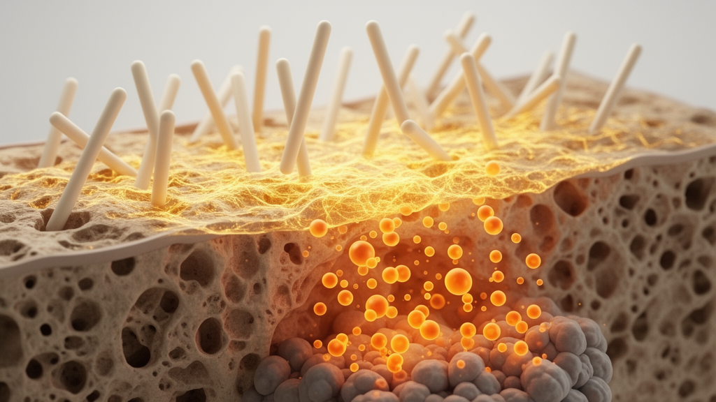
Diabetic patients who suffer serious bone defects face a healing process that is fundamentally broken. High blood sugar floods the injured tissue with reactive oxygen species, sustains chronic inflammation, and accumulates advanced glycation end-products that sabotage the cellular machinery of repair. Fractures heal slowly or not at all, infections escalate quickly, and implants fail at elevated rates. Now, a research team reports a therapy that attacks this hostile microenvironment on several fronts at once, pairing an endogenous anti-inflammatory metabolite with carefully staged doses of heat delivered by a smart hydrogel.
The strategy, described as Immunometabolic–Hyperthermia Therapy, or IMHT, was tested in diabetic rats with infected defects in the femoral condyle. It rests on a dual-functional material: polydopamine nanoparticles loaded with itaconate, embedded in a gelatin–agarose hydrogel. Itaconate is an immunometabolite produced naturally by activated macrophages from the tricarboxylic acid cycle intermediate cis-aconitate, and it has attracted attention for its anti-inflammatory, antioxidant, and antimicrobial properties. Polydopamine, meanwhile, is a strong photothermal agent that converts near-infrared light into heat, giving the hydrogel a built-in heating element.
The therapeutic schedule is deliberately two-phased. On the first day after surgery, the implanted hydrogel is irradiated with an 808 nm near-infrared laser, driving the defect site to roughly 50 °C for ten minutes. This high-temperature phase is the primary antibacterial weapon: in vitro, heat alone visibly disrupted mature Escherichia coli biofilms, thinning and dispersing the dense bacterial architecture, while the itaconate released from the nanoparticles made a further but moderate contribution to killing. From day three onward, the regimen shifts gears, applying repeated sessions of mild hyperthermia at approximately 41 °C every two days for four weeks, alongside continuous itaconate release.
The material itself was engineered with care. Incorporating itaconate during in situ polymerization changed the supramolecular organization of the polydopamine particles, shifting their surface charge from −4.85 mV to nearly neutral and increasing their hydrodynamic diameter to about 540 nanometers, with a stable mesoporous, spherical structure that persisted over fourteen days in water and culture medium. High-performance liquid chromatography confirmed that the nanoparticles released itaconate continuously over twenty-eight days at biologically relevant concentrations, reaching 255.4 micromolar at a particle concentration of 1 milligram per milliliter. The hydrogels showed interconnected pores of 180 to 240 micrometers, a swelling ratio exceeding 3300 percent in the highest-loading formulation, and near-complete degradation over four weeks, with photothermal responsiveness retained even as the matrix broke down.
In cell experiments, the combination therapy showed a striking synergy against the oxidative stress that plagues diabetic tissue. When osteoblast-like MC3T3-E1 cells were challenged with hydrogen peroxide, the combined treatment of itaconate-loaded nanoparticles plus mild hyperthermia reduced intracellular reactive oxygen species to near-healthy levels, preserved mitochondrial membrane potential, and restored alkaline phosphatase activity, mineral deposition, and the expression of osteogenic genes including RUNX2 and osteocalcin to values comparable with cells grown without inflammatory challenge. Neither component alone matched this performance; mild heat by itself offered limited antioxidant protection, while itaconate alone provided only partial mitigation.
Endothelial cells benefited in parallel. In tube formation assays on Matrigel, cells exposed to the combined therapy built robust, intact tubular networks resembling those of unchallenged controls, whereas cells treated with peroxide, plain nanoparticles, or either single therapy formed fragmented vessels. The combination also upregulated the angiogenic genes Vegfa, Pdgfa, and α-SMA, addressing the vascular insufficiency that starves diabetic bone of oxygen and nutrients. Mitochondrial membrane potential in the endothelial cells followed the same protective pattern seen in the osteoblasts.
The immune effects were equally pronounced. In lipopolysaccharide-stimulated macrophages, flow cytometry showed that the combined treatment most strongly suppressed the M1 marker CD86 while raising the M2 marker CD206, and it maximized expression of the anti-inflammatory gene Arg-1 while silencing TNF-α and iNOS. In neutrophils, the therapy suppressed the formation of neutrophil extracellular traps, the web-like DNA structures that amplify diabetic inflammation and damage osteoblasts and endothelial cells. Mitochondrial membrane potential in the macrophages was likewise preserved at near-normal levels only by the combined treatment.
Transcriptomic analysis pointed to a convergent molecular target. Bioinformatic mining of public datasets from itaconate-derivative-treated neutrophils and macrophages, together with new profiling of the nanoparticle-treated cells, repeatedly highlighted the JAK1/STAT1 signaling axis and the MAPK pathway, with STAT1 and then JAK1 emerging as central hub genes. Western blotting confirmed that the combined therapy suppressed agonist-induced phosphorylation of JAK1 and STAT1 more completely than either itaconate or mild heat alone. Meanwhile, the treatment boosted macrophage secretion of fibroblast growth factor-2 to roughly two to three times the level of healthy controls, and mild heat was shown to upregulate heat shock protein 90, which stabilizes phosphorylated FGFR1 and amplifies FGF-2 signaling in target cells. Blocking HSP90 with geldanamycin attenuated this FGFR1 activation, supporting the proposed mechanism in which heat both calms inflammation and potentiates growth factor signaling.
The in vivo results tied the whole program together. In diabetic rats whose femoral defects were seeded with E. coli isolated from infected diabetic bone, the initial high-temperature treatment sharply reduced bacterial counts by day seven, and Giemsa staining found no residual bacterial colonies in the heated groups at six weeks. Micro-computed tomography revealed near-complete defect restoration in the full IMHT group, with significantly improved bone volume fraction, trabecular thickness, trabecular number, and bone mineral density compared with hydrogel implantation or antibacterial heat alone. Histology showed abundant, well-organized new bone, widespread osteocalcin expression, reduced iNOS-positive inflammatory cells, and elevated CD206-positive reparative macrophages. Major organs showed no signs of toxicity, and the 50 °C phase produced only mild local histological changes without osteonecrosis.
The authors caution that important questions remain before such a platform could approach clinical use. The long-term fate of the released nanoparticles, including their tissue retention, biodistribution, and clearance kinetics in the diabetic microenvironment, requires further quantification, and the study was conducted in a rat model over a one-month treatment window. Still, the work demonstrates a coherent design principle: rather than treating infection, inflammation, and failed regeneration as separate problems, a single material can sequence antibacterial heat, sustained immunometabolic reprogramming, and regenerative thermal activation in time, allowing each intervention to prepare the ground for the next.
Subject of Research: Immunometabolic–hyperthermia nanotherapy for regenerating infected diabetic bone defects
Article Title: Spatiotemporally coordinated immunometabolic-hyperthermia therapy for diabetic bone defect regeneration
Article References: Wang, L., Peng, X., Zheng, L., Liu, Y., Chen, L., Li, X., & Wang, Z. (2026). Spatiotemporally coordinated immunometabolic-hyperthermia therapy for diabetic bone defect regeneration. Materials Today Bio, 41, Article 103767. https://doi.org/10.1016/j.mtbio.2026.103767
Image Credits: AI Generated
DOI: 10.1016/j.mtbio.2026.103767
Keywords: diabetic bone defects, itaconate, polydopamine nanoparticles, mild hyperthermia, macrophage polarization, JAK1/STAT1, FGF-2, HSP90, photothermal therapy, biofilm disruption, bone tissue engineering, hydrogel
Cite Scienmag News
Denise Maddox. (October 23, 2026). Heat and Itaconate Combine to Rebuild Bone in Diabetes. Scienmag. https://scienmag.com/heat-and-itaconate-combine-to-rebuild-bone-in-diabetes/
Denise Maddox. "Heat and Itaconate Combine to Rebuild Bone in Diabetes." Scienmag, 23 October 2026, https://scienmag.com/heat-and-itaconate-combine-to-rebuild-bone-in-diabetes/. Accessed 23 October 2026.
Denise Maddox. "Heat and Itaconate Combine to Rebuild Bone in Diabetes." Scienmag. October 23, 2026. https://scienmag.com/heat-and-itaconate-combine-to-rebuild-bone-in-diabetes/


