


Ovarian cancer has long been one of medicine’s most unforgiving diagnoses, and the reason often comes down to a single factor: time. When the disease is detected in its early stages, the five-year survival rate can climb above 90 percent. But when it is caught late, at stage 3 or 4, that figure falls to less than half. The brutal arithmetic of these numbers has driven researchers for decades to search for tools that can spot the disease before it spreads, and a new piece of engineering from the Massachusetts Institute of Technology may bring that goal significantly closer.
Researchers in the laboratory of Professor Kripa Varanasi at MIT, working with collaborators at MIT and Johns Hopkins University, have developed a handheld device capable of gently collecting living cells from precise locations within tissue samples. Described in the journal Device, the system is designed to sample the fallopian tubes, where roughly two decades of research have revealed that many cases of high-grade serous ovarian cancer, the most common form of the disease, actually begin. Because precursor lesions in the fallopian tubes can be microscopic and extraordinarily difficult to sample, they have remained largely invisible to conventional diagnostic workflows.
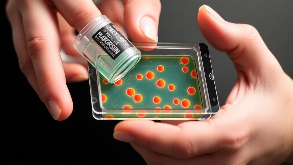
The technical concept behind the device is elegantly simple, even if its execution required careful engineering. A 3D-printed microfluidic chip is pressed against the surface of freshly excised tissue, where one syringe creates a vacuum that seals the device against the specimen, ensuring that nothing leaks during operation. A second syringe then pushes fluid through a small microfluidic channel, producing a flow that applies shear stress parallel to the tissue surface. Rather than cutting or scraping cells away with a scalpel or brush, the flowing liquid gently detaches living cells from tiny, targeted sections of tissue, leaving the surrounding structure intact for conventional pathological examination.
“We came up with this device where one syringe creates a vacuum that holds it against the tissue, and a second syringe pushes liquid through it,” says Bert Vandereydt, an MIT postdoc and co-first author of the study. “The vacuum creates a seal, so nothing leaks, and then we locally apply what is basically a microfluidic chip on the tissue that gently shears the cells off.” The insight emerged from the lab’s deep experience with fluid-surface interfaces, and from conversations with surgeons at Johns Hopkins who observed that certain cells were loose enough to come off during routine washing and other procedures in the operating room.
The significance of collecting cells alive, rather than fixed in preservative, cannot be overstated. The traditional approach to examining removed fallopian tubes involves placing the tissue in a chemical preservative that maintains structure but kills the cells, making it impossible to grow them in culture. A pathologist must then painstakingly search thin sections under a microscope for cancerous or precancerous regions, a process that Varanasi describes as time-consuming and destructive to the cells. With the new technique, researchers can not only see what cells look like but recover them viable, cultivate them, grow them into organoids, and build living models of disease that show how cells from a specific patient behave.
In their study, the researchers demonstrated that the shear stress applied by the device can be tuned to different levels, and they compared their approach against other cell-collection workflows. The cells harvested with the fluidic method remained viable and grew in culture far more readily than those detached by conventional means. The tunability extends across disease types as well. Loose prostate cancer cells detach under stress as low as one pascal, whereas bone cancer cells proved far more tenaciously adherent, with only a few cells releasing even under five pascals of applied stress. As Vandereydt puts it, the approach is agnostic to the disease, which suggests broad utility well beyond ovarian cancer.
Proving the technology in real clinical conditions required an unusual level of dedication. The team tested the device on fresh human fallopian tube samples shipped from collaborators at Johns Hopkins, which meant staying on call for deliveries that could arrive at unpredictable times. “Sometimes, we’d get an email from our collaborators at 11 p.m. saying ‘There are two fallopian tubes coming tomorrow,'” Vandereydt recalls. After each experiment, the samples were returned for conventional pathology, ensuring that nothing was lost from the diagnostic record. From those fresh specimens, the team successfully collected living cells and converted them into organoids, the crucial building blocks for testing, disease modeling, and eventually personalized treatment development.
The project traces its origins to a fortunate convergence of clinical and engineering expertise. Rebecca Stone, a gynecologic oncologist at the Johns Hopkins University School of Medicine, has long advocated for risk-reducing fallopian tube removal for certain women at elevated risk, a procedure that can be performed after childbearing years while largely preserving normal hormone production. When Angela Belcher, MIT’s James Mason Crafts Professor, introduced Stone to Varanasi, the collaboration was born, funded by Break Through Cancer, a foundation that assembles interdisciplinary teams to attack the most stubborn problems in oncology. “This is exactly the kind of problem that benefits from bringing clinicians and engineers together,” Stone says. “We understand the clinical need, while the MIT team brings a very different perspective from fluid mechanics and engineering.”
The road ahead involves two distinct phases. In the near term, the researchers plan to apply the device to tissue that has already been removed from the body, a pathway that offers easier regulatory approval and could transform routine histopathology by allowing living cells to be harvested while the specimen is preserved for standard examination. Stone notes that integrating the technology into existing pathology workflows could create a powerful new way to study carcinogenesis and fundamental biology directly from human tissue. In parallel, Belcher’s group is developing optical approaches to identify suspicious regions of tissue, and the cell-collection device could then extract living cells from exactly those flagged locations. The longer-term ambition is bolder still: adapting the device to swab samples inside patients directly, enabling easier testing and earlier detection without surgery.
What makes this work resonate far beyond a single laboratory is the vision it points toward for cancer medicine as a whole. If clinicians can harvest living cells from specific, suspicious regions without destroying them, then diagnostics could evolve from static snapshots of preserved tissue into dynamic studies of living disease. Organoids grown from a patient’s own cells could be exposed to different therapies in the laboratory, revealing which treatments work before a patient receives a single dose, a genuine step toward personalized medicine. Varanasi credits Break Through Cancer with making the collaboration possible, noting that teams working on pancreatic cancer, brain cancer, leukemia, and other diseases all expressed how valuable better methods for obtaining living cells from specific tissue regions would be. For the researchers, the ultimate measure of success is human: “If this work can ultimately help women by enabling earlier detection of ovarian cancer, I would find that incredibly fulfilling,” Varanasi says. “That is really what motivates us — taking the science and engineering we develop in the lab and using it to make a difference in people’s lives.”
Subject of Research: A handheld microfluidic shear-stress device for collecting viable cells from fallopian tube tissue to improve early cancer detection
Article Title: New cell-collection device could improve early cancer detection
Article References: New cell-collection device could improve early cancer detection. (n.d.). Original publication
Image Credits: AI Generated
DOI: Not provided
Keywords: ovarian cancer, microfluidics, fallopian tubes, early detection, organoids, living cells, personalized medicine, MIT, shear stress, cancer diagnostics, pathology, biomedical engineering
Cite Scienmag News
Nathaniel Bowman. (October 15, 2026). Handheld Microfluidic Device Collects Living Cells to Catch Cancer Earlier. Scienmag. https://scienmag.com/handheld-microfluidic-device-collects-living-cells-to-catch-cancer-earlier/
Nathaniel Bowman. "Handheld Microfluidic Device Collects Living Cells to Catch Cancer Earlier." Scienmag, 15 October 2026, https://scienmag.com/handheld-microfluidic-device-collects-living-cells-to-catch-cancer-earlier/. Accessed 15 October 2026.
Nathaniel Bowman. "Handheld Microfluidic Device Collects Living Cells to Catch Cancer Earlier." Scienmag. October 15, 2026. https://scienmag.com/handheld-microfluidic-device-collects-living-cells-to-catch-cancer-earlier/


