


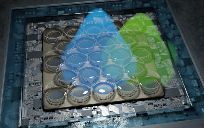
What does the inside of a cell really look like? In the past, standard microscopes were limited in how well they could answer this question. Now, researchers from the Universities of Göttingen and Oxford, in collaboration with the University Medical Center Göttingen (UMG), have succeeded in developing a microscope with resolutions better than five nanometres (five billionths of a metre). This is roughly equivalent to the width of a hair split into 10,000 strands. Their new method was published in Nature Photonics.

Credit: Alexey Chizhik, Göttingen University
What does the inside of a cell really look like? In the past, standard microscopes were limited in how well they could answer this question. Now, researchers from the Universities of Göttingen and Oxford, in collaboration with the University Medical Center Göttingen (UMG), have succeeded in developing a microscope with resolutions better than five nanometres (five billionths of a metre). This is roughly equivalent to the width of a hair split into 10,000 strands. Their new method was published in Nature Photonics.
Many structures in cells are so small that standard microscopes can only produce fragmented images. Their resolution only begins at around 200 nanometres. However, human cells for instance contain a kind of scaffold of fine tubes that are only around seven nanometres wide. The synaptic cleft, meaning the distance between two nerve cells or between a nerve cell and a muscle cell, is just 10 to 50 nanometres – too small for conventional microscopes. The new microscope, which researchers at the University of Göttingen have helped to develop, promises much richer information. It benefits from a resolution better than five nanometres, enabling it to capture even the tiniest cell structures. It is difficult to imagine something so tiny, but if we were to compare one nanometre with one metre, it would be the equivalent of comparing the diameter of a hazelnut with the diameter of the Earth.
This type of microscope is known as a fluorescence microscope. Their function relies on “single-molecule localization microscopy”, in which individual fluorescent molecules in a sample are switched on and off and their individual positions are then determined very precisely. The entire structure of the sample can then be modelled from the positions of these molecules. The current process enables resolutions of around 10 to 20 nanometres. Professor Jörg Enderlein’s research group at the University of Göttingen’s Faculty of Physics has now been able to double this resolution again – with the help of a highly sensitive detector and special data analysis. This means that even the tiniest details of protein organization in the connecting area between two nerve cells can be very precisely revealed.
“This newly developed technology is a milestone in the field of high-resolution microscopy. It not only offers resolutions in the single-digit nanometre range, but it is also particularly cost-effective and easy to use compared to other methods,” explains Enderlein. The scientists also developed an open-source software package for data processing in the course of publishing their findings. This means that this type of microscopy will be available to a wide range of specialists in the future.
Original publication: Jörg Enderlein et al. “Doubling the resolution of fluorescence-lifetime single-molecule localization microscopy with image scanning microscopy”. Nature Photonics 2024. DOI: 10.1038/s41566-024-01481-4
Contact:
Professor Jörg Enderlein
University of Göttingen
Faculty of Physics – Biophysics and complex systems
Friedrich Hund Platz 1, 37077 Göttingen, Germany
Tel: +49 (0)551 39 26908
Email: joerg.enderlein@phys.uni-goettingen.de
What does the inside of a cell really look like? In the past, standard microscopes were limited in how well they could answer this question. Now, researchers from the Universities of Göttingen and Oxford, in collaboration with the University Medical Center Göttingen (UMG), have succeeded in developing a microscope with resolutions better than five nanometres (five billionths of a metre). This is roughly equivalent to the width of a hair split into 10,000 strands. Their new method was published in Nature Photonics.
Many structures in cells are so small that standard microscopes can only produce fragmented images. Their resolution only begins at around 200 nanometres. However, human cells for instance contain a kind of scaffold of fine tubes that are only around seven nanometres wide. The synaptic cleft, meaning the distance between two nerve cells or between a nerve cell and a muscle cell, is just 10 to 50 nanometres – too small for conventional microscopes. The new microscope, which researchers at the University of Göttingen have helped to develop, promises much richer information. It benefits from a resolution better than five nanometres, enabling it to capture even the tiniest cell structures. It is difficult to imagine something so tiny, but if we were to compare one nanometre with one metre, it would be the equivalent of comparing the diameter of a hazelnut with the diameter of the Earth.
This type of microscope is known as a fluorescence microscope. Their function relies on “single-molecule localization microscopy”, in which individual fluorescent molecules in a sample are switched on and off and their individual positions are then determined very precisely. The entire structure of the sample can then be modelled from the positions of these molecules. The current process enables resolutions of around 10 to 20 nanometres. Professor Jörg Enderlein’s research group at the University of Göttingen’s Faculty of Physics has now been able to double this resolution again – with the help of a highly sensitive detector and special data analysis. This means that even the tiniest details of protein organization in the connecting area between two nerve cells can be very precisely revealed.
“This newly developed technology is a milestone in the field of high-resolution microscopy. It not only offers resolutions in the single-digit nanometre range, but it is also particularly cost-effective and easy to use compared to other methods,” explains Enderlein. The scientists also developed an open-source software package for data processing in the course of publishing their findings. This means that this type of microscopy will be available to a wide range of specialists in the future.
Original publication: Jörg Enderlein et al. “Doubling the resolution of fluorescence-lifetime single-molecule localization microscopy with image scanning microscopy”. Nature Photonics 2024. DOI: 10.1038/s41566-024-01481-4
Contact:
Professor Jörg Enderlein
University of Göttingen
Faculty of Physics – Biophysics and complex systems
Friedrich Hund Platz 1, 37077 Göttingen, Germany
Tel: +49 (0)551 39 26908
Email: joerg.enderlein@phys.uni-goettingen.de
Journal
Nature Photonics
Method of Research
Experimental study
Subject of Research
Not applicable
Article Title
Doubling the resolution of fluorescence-lifetime single-molecule localization microscopy with image scanning microscopy
Article Publication Date
2-Aug-2024
Cite Scienmag News
Denise Maddox. (August 7, 2024). Glimpse into the nanoworld: microscope reveals tiniest cell processes. Scienmag. https://scienmag.com/glimpse-into-the-nanoworld-microscope-reveals-tiniest-cell-processes/
Denise Maddox. "Glimpse into the nanoworld: microscope reveals tiniest cell processes." Scienmag, 7 August 2024, https://scienmag.com/glimpse-into-the-nanoworld-microscope-reveals-tiniest-cell-processes/. Accessed 4 September 2026.
Denise Maddox. "Glimpse into the nanoworld: microscope reveals tiniest cell processes." Scienmag. August 7, 2024. https://scienmag.com/glimpse-into-the-nanoworld-microscope-reveals-tiniest-cell-processes/

