


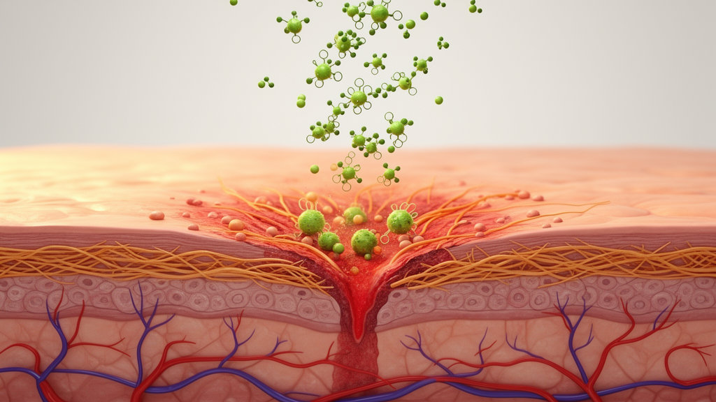
Deep or complex wounds that refuse to heal remain one of medicine’s most stubborn problems. According to a comprehensive review published in the Journal of Cellular and Molecular Medicine, an estimated 1% to 2% of the global population suffers from refractory wounds, a figure climbing steadily alongside ageing populations and the rising burden of diabetes. Diabetic foot ulcers are especially punishing: their annual recurrence rate exceeds 30%, and conventional tools—debridement, antibiotics, standard dressings, costly recombinant growth factors and skin grafts—often deliver prolonged healing cycles, drug resistance, scarring and other complications. Against this backdrop, researchers are taking a hard scientific look at an ancient remedy: the saponins of Panax notoginseng, a herb long prized in traditional Chinese medicine, whose active compounds can now be purified to levels exceeding 90%.
The review, led by Zhifeng Wang and colleagues, maps how Panax notoginseng saponins (PNS) act on all four sequential phases of skin wound repair: haemostasis, inflammation, proliferation and remodelling. The mixture’s principal active monomers are ginsenosides Rg1 and Rb1, supported by minor components such as ginsenosides Rd and Re and notoginsenoside R1. Rather than a single blunt instrument, PNS behaves like a coordinated molecular toolkit, modulating the inflammatory microenvironment, accelerating cell proliferation and migration, driving angiogenesis and suppressing scar formation through multiple signalling pathways at once.
In the inflammatory phase, the first act of healing, moderate inflammation is useful—it clears debris and necrotic tissue—but excessive or persistent inflammation derails repair. In cell and animal studies, total PNS significantly downregulates pro-inflammatory cytokines including IL-6, IL-8 and TNF-α, reduces infiltration of neutrophils and macrophages, and helps the wound transition smoothly into the proliferative phase. The two flagship monomers divide the labour. Ginsenoside Rg1 is the core driver of pro-inflammatory suppression, targeting the NF-κB pathway and dose-dependently reducing TNF-α and IL-1β transcription. Ginsenoside Rb1 more specifically inhibits IL-6 and IL-8 and dampens mast cell degranulation, curbing histamine and tryptase release. At equivalent doses, the total saponin mixture outperforms either monomer alone, with no apparent dose-dependent toxicity—a striking illustration of multi-monomer synergy.
The molecular plumbing runs deeper still. Total PNS simultaneously targets the two central inflammatory signalling cascades: it blocks NF-κB activation by inhibiting degradation of IκBα and preventing nuclear translocation of the NF-κB p65 subunit, while also suppressing phosphorylation of ERK1/2, p38 and JNK in the MAPK pathway. Rb1 adds a layer of finesse, bidirectionally regulating p38 and JNK—inhibiting excessive JNK phosphorylation while moderately activating ERK1/2 to promote repair. Crucially, PNS also upregulates the anti-inflammatory cytokine IL-10 and pushes macrophages from the pro-inflammatory M1 phenotype toward the reparative M2 state. Rb1 is the star here, triggering M2 polarization more than three times as strongly as Rg1, upregulating M2 markers CD206 and Arg-1 and prompting M2 macrophages to secrete reparative factors such as TGF-β1 and VEGF. Yet the picture is context-dependent: in tumour models, PNS has been reported to push macrophages toward M1 instead, suggesting that the balance between Rg1 and Rb1—and the local microenvironment—dictates the outcome.
When healing shifts into the proliferative phase, PNS again shows two-pronged precision. Keratinocytes, the workhorses of re-epithelialization, are Rg1’s domain: total PNS activates the PI3K/AKT-ERK1/2 axis, pushes HaCaT keratinocytes from the G0/G1 phase into S phase, increases PCNA-positive cells and upregulates MMP-2/9 activity to clear spatial barriers to migration. Rb1, by contrast, barely touches keratinocytes but is the key regulator of fibroblasts, the cells that build granulation tissue. Through functional modulation of the TGF-β1 type II receptor, Rb1 bidirectionally governs fibroblast behaviour—promoting proliferation and collagen synthesis early, restraining excessive activation later, and maintaining the physiological 3:1 ratio of type I to type III collagen. Notably, in diabetic wound models, total PNS upregulates type III collagen more than type I, potentially deviating from that physiological ratio, hinting that other monomers exert non-specific effects that still need untangling.
Angiogenesis—the growth of new blood vessels that supply oxygen, nutrients and cytokines—is where the monomer partnership becomes most dramatic. Rg1 dose-dependently enhances VEGF expression via the HIF-1α/VEGF pathway, driving endothelial cell proliferation, migration and tube formation, and activates the PI3K/AKT pathway to raise anti-apoptotic Bcl-2 in endothelial cells. Rb1, which does not upregulate VEGF, instead boosts bFGF to promote basement membrane formation and vascular maturation, and antagonizes the VEGF overexpression caused by high-concentration Rg1—reducing risks of vascular leakage and aberrant proliferation. Total PNS also activates the JAK2-STAT3 pathway through Rb1 and inhibits platelet adhesion and aggregation to improve local circulation, with Rg1 carrying the stronger anticoagulant effect. One caveat deserves emphasis: the pro-angiogenic effect is microenvironment-dependent, significant under normoxic conditions but markedly weakened in the severely hypoxic milieu of chronic wounds—a puzzle tied to Rg1’s HIF-1α regulation that remains unresolved.
The remodelling phase, where scars are won or lost, showcases PNS’s anti-fibrotic credentials. A central mechanism of scar formation is the transition of fibroblasts into myofibroblasts, governed by the TGF-β1/Smad2/3 pathway. Total PNS blocks TGF-β1 receptor binding and inhibits Smad2/3 phosphorylation and nuclear translocation, downregulating α-SMA and easing scar hyperplasia—effects attributed mainly to Rb1. Meanwhile Rg1 fights oxidative stress, boosting activities of superoxide dismutase and glutathione peroxidase while reducing malondialdehyde accumulation, thereby protecting fibroblast and endothelial function during remodelling. In mouse models of post-traumatic scarring, topical PNS produced significantly smaller scar areas than saline controls, with improved tissue quality and no observable local adverse reactions. In human hypertrophic scar fibroblasts, PNS treatment reduced type I/III collagen synthesis, migration and α-SMA expression.
Translating these mechanisms to the bedside has demanded creative delivery engineering, because oral PNS is degraded by gastric acid and intestinal enzymes, while conventional topicals penetrate poorly and release drug unevenly. The review catalogues a new generation of carriers: PLGA nanoparticles with degradation cycles tunable from one to six months to match the healing timeline; chitosan nanoparticles offering intrinsic antibacterial, hemostatic and bioadhesive properties, though with a substantial 30%–50% burst release within 24 hours; chitosan-based hydrogels whose porous, extracellular-matrix-mimicking scaffolds support cell adhesion; thermosensitive PNIPAM hydrogels that stay liquid at room temperature and gel in situ at body temperature, releasing drug steadily for one to two weeks; and PCL electrospun fibrous membranes whose nanoscale networks resemble natural skin ECM and can be co-loaded with silver ions or antimicrobial peptides. Most futuristic of all are intelligent dressings: pH-responsive systems that release more drug as wound inflammation raises pH, and near-infrared-responsive nanofibers using graphene or gold nanoparticles to trigger pulsed, on-demand release—though the latter requires specialized equipment and raises unresolved biosafety questions.
Preclinical results across wound types are encouraging. PNS creams shortened healing time and increased tensile strength in rat incision models; PNS composite hydrogels significantly raised healing rates in diabetic foot ulcer models, improving granulation tissue regularity and regenerating hair follicles and sebaceous glands; and in superficial second-degree burns, PNS formulations upregulated PDGF-BB/PDGFR-β, accelerating granulation and epithelial repair. Early clinical exploration suggests topical PNS accelerates healing of surgical incisions and burns with favourable skin safety and no obvious irritation or allergic reactions. But the evidence hierarchy remains sobering: most mechanistic data sit at the in vitro level, animal findings form the preclinical bridge, and human evidence comes from single-centre, small-sample studies that cannot yet support high-grade clinical recommendations.
The authors are candid about the gaps. No study has yet used chemoproteomic target-deconvolution techniques such as surface plasmon resonance, microscale thermophoresis or cellular thermal shift assays to confirm the direct binding targets of PNS monomers in skin-wound systems—current evidence relies on downstream phosphorylation readouts, and several key conclusions were extrapolated from pulmonary fibrosis or oral mucosal models rather than skin. Extraction processes add another variable: ultrasonic-assisted extraction enriches Rg1 and Rb1, supercritical CO2 extraction maximizes purity but under-recovers notoginsenoside R1, and shifting Rg1:Rb1 ratios can alter—or even reverse—the direction of synergistic effects, potentially explaining contradictory results across studies. The proposed path forward pairs total PNS for full-cycle routine treatment with staged, precise monomer intervention: Rg1 for delayed epithelialization, Rb1 for high-scar-risk remodelling, and sequenced administration for diabetic ulcers. With single-cell sequencing, spatial transcriptomics, 3D bioprinted skin, dual-responsive dressings and mesenchymal stem cell co-delivery on the roadmap, this ancient saponin mixture may be moving from empirical tradition toward precision regenerative medicine—provided multicentre randomized trials and unified quality-control standards can finally catch up with the chemistry.
Subject of Research: Mechanisms and delivery systems of Panax notoginseng saponins in skin wound repair
Article Title: Application of Panax notoginseng Saponins in Skin Wound Repair: Mechanisms and Research Progress
Article References: Wang, Z., Shi, T., Tian, D., Li, R., Yao, T., Lan, J., Xi, Y., Wang, R., & Wang, J. (2026). Application of Panax notoginseng Saponins in Skin Wound Repair: Mechanisms and Research Progress. Journal of Cellular and Molecular Medicine, 30(20), Article e71381. https://doi.org/10.1111/jcmm.71381
Image Credits: AI Generated
DOI: 10.1111/jcmm.71381
Keywords: Panax notoginseng, wound healing, ginsenoside Rg1, ginsenoside Rb1, saponins, angiogenesis, scar formation, macrophage polarization, nanocarriers, hydrogels, smart dressings, diabetic foot ulcers
Cite Scienmag News
Drew Townsend. (October 16, 2026). From Ancient Herb to Smart Dressing: How Notoginseng Saponins Rewire Every Stage of Wound Healing. Scienmag. https://scienmag.com/from-ancient-herb-to-smart-dressing-how-notoginseng-saponins-rewire-every-stage-of-wound-healing/
Drew Townsend. "From Ancient Herb to Smart Dressing: How Notoginseng Saponins Rewire Every Stage of Wound Healing." Scienmag, 16 October 2026, https://scienmag.com/from-ancient-herb-to-smart-dressing-how-notoginseng-saponins-rewire-every-stage-of-wound-healing/. Accessed 16 October 2026.
Drew Townsend. "From Ancient Herb to Smart Dressing: How Notoginseng Saponins Rewire Every Stage of Wound Healing." Scienmag. October 16, 2026. https://scienmag.com/from-ancient-herb-to-smart-dressing-how-notoginseng-saponins-rewire-every-stage-of-wound-healing/


