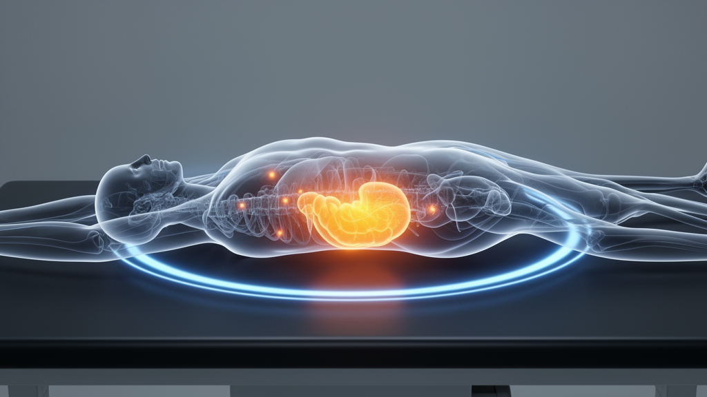
One of the most feared events in stomach cancer is not the primary tumor itself but its silent spread to the peritoneum, the thin membrane lining the abdominal cavity. Peritoneal metastasis marks a turning point in the disease: once cancer cells seed this delicate lining, curative surgery is usually off the table and survival drops sharply. The problem for doctors is that these seedings are notoriously hard to see. Conventional imaging with computed tomography often misses small peritoneal deposits, and the current gold standard for detection, staging laparoscopy with peritoneal washing cytology, is an invasive surgical procedure. A new study published in BMC Medical Imaging by Qipeng Yuan and colleagues at Shandong Cancer Hospital and Institute now offers one of the most rigorous assessments to date of a promising molecular imaging technique, fibroblast activation protein inhibitor PET/CT, known as FAPI PET/CT, for detecting this hidden spread. The results are a study in contrasts: striking specificity, but sensitivity that falls short of what clinicians would hope for.
The technology behind FAPI imaging represents a significant departure from the fluorodeoxyglucose, or FDG, tracers that have dominated nuclear medicine for decades. Rather than tracking glucose metabolism, FAPI tracers home in on fibroblast activation protein, a molecule abundantly expressed by cancer-associated fibroblasts, the stromal cells that build the tumor’s supporting scaffold. Because these fibroblasts are woven throughout the tumor microenvironment, FAPI tracers can light up tumors with remarkably high target-to-background contrast. In principle, that brightness against a quiet background should make even small peritoneal implants visible on a scan. The Shandong team used a tracer called [18F]AlF-NOTA-FAPI-04, a fluorine-18 labeled compound that can be produced in standard radiochemistry facilities, an advantage over gallium-68 based FAPI agents that require a generator on site.
The study had two complementary components. First, the researchers ran an exploratory prospective cohort study in which patients with gastric cancer underwent FAPI PET/CT before reference testing. The reference standard was deliberately stringent: a laparoscopy-based composite that combined direct visual inspection of the peritoneal cavity, peritoneal washing cytology, and histopathological examination of any suspicious lesions. This matters because many earlier studies of FAPI imaging relied on loosely defined reference standards, often a multidisciplinary team’s consensus judgment that incorporated the imaging itself, creating a risk of circular reasoning that inflates apparent accuracy. By anchoring their prospective cohort to laparoscopy, the investigators aimed to measure the scan’s true performance against the most credible benchmark available short of full laparotomy.
The prospective results were sobering on one front and encouraging on another. FAPI PET/CT achieved a sensitivity of just 0.40, with a 95 percent confidence interval spanning 0.12 to 0.77, meaning the scan missed the majority of patients who actually had peritoneal metastasis in this cohort. Yet the specificity was a perfect 1.00, with a confidence interval of 0.72 to 1.00: every positive scan corresponded to genuine disease, and there were no false alarms. Overall accuracy reached 0.80, with a positive predictive value of 1.00 and a negative predictive value of 0.77. The authors are candid about the limitations of these estimates. The cohort was small, the confidence intervals are wide, and the point estimates should be interpreted cautiously. A sensitivity of 0.40 in a small sample could plausibly reflect either a genuinely poor ability to detect subtle peritoneal seeding or statistical uncertainty around a higher true value; the data cannot yet distinguish between those possibilities.
To place their cohort findings in context, the team conducted a systematic review and meta-analysis, searching PubMed, Embase, the Cochrane Library, and Web of Science through July 10, 2026. They included studies that evaluated FAPI-04 PET/CT for peritoneal metastasis in gastric cancer and reported per-patient diagnostic data suitable for constructing two-by-two tables of true positives, false positives, true negatives, and false negatives. Methodological quality was appraised with QUADAS-2, the standard tool for diagnostic accuracy studies, and pooled sensitivity and specificity were jointly estimated using a bivariate random-effects model, which accounts for the inherent trade-off between these two metrics. The reporting followed PRISMA-DTA and STARD guidelines, and the review was registered prospectively, signaling a commitment to transparency unusual in this literature.
The meta-analysis revealed how much the choice of reference standard shapes the apparent performance of FAPI PET/CT. In the primary analysis, restricted to datasets that used a laparoscopy-based composite reference standard, pooled sensitivity was 0.66 and pooled specificity was 0.90. When the researchers analyzed studies that used multidisciplinary team-based assessment as the reference, the numbers jumped dramatically: sensitivity of 0.92 and specificity of 0.98. Across all eleven datasets eligible for bivariate pooling, regardless of reference standard, the pooled figures were 0.86 sensitivity and 0.96 specificity. The pattern is instructive. When the truth is defined by objective, invasive testing, the scan looks considerably less sensitive than when the truth is defined partly by the imaging itself and clinical judgment. This discrepancy does not mean the higher numbers are fraudulent, but it does illustrate a well-known pitfall in diagnostic research: incorporation bias, where the test under evaluation contributes to the very standard against which it is judged.
What does this mean for patients with gastric cancer? The clinical stakes are high. Peritoneal metastasis is present in a meaningful fraction of patients at initial diagnosis, including a subset with so-called CY1P0 disease, in which cancer cells are found in peritoneal washings even though no visible implants exist. Identifying these patients before surgery spares them a major operation that cannot cure them and redirects them toward systemic therapy. A noninvasive scan that could reliably replace or triage staging laparoscopy would be transformative. FAPI PET/CT’s perfect specificity in the prospective cohort suggests that a positive scan can be trusted, potentially allowing clinicians to proceed directly to palliative systemic treatment without confirmatory laparoscopy in selected cases. The weak sensitivity, however, means a negative scan cannot rule out peritoneal disease, and laparoscopy would remain necessary to exclude spread with confidence.
The biological explanation for missed lesions is worth considering. Peritoneal metastases in gastric cancer are often tiny, sometimes only a few millimeters, and may consist of scattered malignant cells within the peritoneal fluid rather than organized tumor nodules. A PET scanner’s ability to resolve a lesion depends on its size, the density of FAP-expressing fibroblasts within it, and the background activity of the tracer. Microscopic or cytology-positive disease without macroscopic deposits may simply fall below the detection threshold of any current imaging modality. Conversely, the stroma-rich nature of gastric tumors means that when a deposit is large enough and fibroblast-rich enough, FAPI PET/CT can detect it with exceptional clarity, which is precisely why specificity remains so high. The scan, in other words, is very good at confirming disease it can see, but blind to disease it cannot resolve.
The authors also explored sources of heterogeneity across the pooled studies using univariable meta-regression, an approach that tests whether factors such as reference standard type explain the variation in reported accuracy between studies. The QUADAS-2 assessment and the sensitivity analyses comparing laparoscopy-anchored with MDT-anchored datasets effectively serve the same interpretive purpose, and the divergence between those analyses is itself the clearest signal of where heterogeneity originates. The work was supported by the National Natural Science Foundation of China and a Collaborative Academic Innovation Project of Shandong Cancer Hospital, and it was conducted under the Declaration of Helsinki with institutional ethics approval and written informed consent from all participants.
The bottom line is a measured but genuinely useful advance. FAPI PET/CT is not yet ready to replace staging laparoscopy for gastric cancer, and the Shandong team says so explicitly, calling for large-scale, well-designed prospective studies to validate diagnostic performance and clinical utility. But the technique’s near-perfect specificity, its practical fluorine-18 chemistry, and the growing body of pooled evidence suggesting sensitivity in the range of 0.66 to 0.86 depending on context make it a serious candidate for integration into staging algorithms, most plausibly as a complement to rather than a substitute for laparoscopy. For a disease in which the difference between a curable and an incurable operation often hinges on detecting invisible peritoneal seeds, even incremental gains in noninvasive detection carry real weight. The study, published open access under DOI 10.1186/s12880-026-02884-1, sets a methodological benchmark for how such imaging tests should be evaluated: prospectively, against invasive reference standards, and with the honesty to report the sensitivity numbers that matter most to patients.
Subject of Research: Diagnostic accuracy of FAPI PET/CT for detecting peritoneal metastasis in gastric cancer
Article Title: Diagnostic performance of FAPI PET/CT for detecting peritoneal metastasis in gastric cancer: an exploratory prospective study and systematic review with meta-analysis
Article References: Diagnostic performance of FAPI PET/CT for detecting peritoneal metastasis in gastric cancer: an exploratory prospective study and systematic review with meta-analysis. (n.d.). https://doi.org/10.1186/s12880-026-02884-1
Image Credits: AI Generated
DOI: 10.1186/s12880-026-02884-1
Keywords: FAPI PET/CT, peritoneal metastasis, gastric cancer, molecular imaging, staging laparoscopy, fibroblast activation protein, meta-analysis, diagnostic accuracy, nuclear medicine, cancer-associated fibroblasts, BMC Medical Imaging, tumor stroma
Cite Scienmag News
Nathaniel Bowman. (October 15, 2026). FAPI PET/CT Shows Promise but Limits in Spotting Hidden Stomach Cancer Spread. Scienmag. https://scienmag.com/fapi-pet-ct-shows-promise-but-limits-in-spotting-hidden-stomach-cancer-spread/
Nathaniel Bowman. "FAPI PET/CT Shows Promise but Limits in Spotting Hidden Stomach Cancer Spread." Scienmag, 15 October 2026, https://scienmag.com/fapi-pet-ct-shows-promise-but-limits-in-spotting-hidden-stomach-cancer-spread/. Accessed 15 October 2026.
Nathaniel Bowman. "FAPI PET/CT Shows Promise but Limits in Spotting Hidden Stomach Cancer Spread." Scienmag. October 15, 2026. https://scienmag.com/fapi-pet-ct-shows-promise-but-limits-in-spotting-hidden-stomach-cancer-spread/