


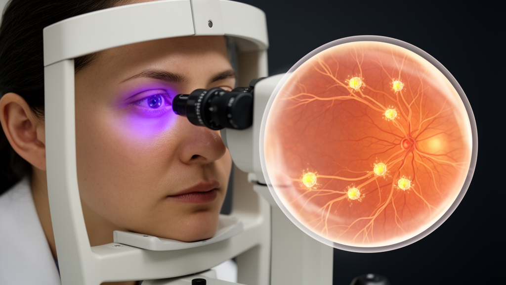
In a finding that could reshape how scientists think about psychiatric illness, researchers in Turkey have used an ophthalmic imaging technique to detect striking alterations in the nerves and immune cells of the eyes of people with obsessive-compulsive disorder. The study, conducted at Gazi University in Ankara and published in BMC Psychiatry, is the first to apply corneal confocal microscopy to OCD, and it reports that patients who had never taken psychiatric medication showed a dramatic increase in corneal immune cells alongside a measurable loss of the fine nerve fibers that weave through the clear front surface of the eye. Because these structures can be imaged in minutes without so much as an eye drop, the work raises the possibility that a routine optical scan might one day offer an objective, biological window into a disorder that is currently diagnosed almost entirely through conversation and questionnaires.
Obsessive-compulsive disorder affects roughly one to two percent of the population and is characterized by intrusive, distressing thoughts and repetitive behaviors performed to neutralize the anxiety they provoke. For decades the condition was understood primarily through psychological and neural circuit models, but a growing body of research has implicated inflammatory and immunological mechanisms as well. Studies have reported altered levels of circulating cytokines, markers of oxidative stress, and immune cell activity in OCD, and some patients show worsening of symptoms after streptococcal infections, hinting that the immune system may participate in the disorder in ways that standard brain imaging cannot easily capture. The problem has always been measurement: directly sampling neuroinflammation in a living brain requires invasive procedures that are impossible to justify in routine research.
The cornea offers an elegant workaround. It is the most densely innervated tissue in the human body, supplied by thousands of slender nerve fibers that branch into a delicate mesh just beneath its surface, known as the subbasal nerve plexus. Critically, this living nerve network can be visualized noninvasively with corneal confocal microscopy, a technique that uses a low-power laser to capture high-resolution images of the cornea at the level of single cells. The method has already proven its worth in other fields: it is widely used to detect the small-fiber nerve damage of diabetic neuropathy, often before symptoms appear, and it has revealed corneal nerve loss in conditions ranging from multiple sclerosis to Parkinson’s disease and dementia. Alongside the nerves, the cornea also hosts resident immune sentinels called dendritic cells, which normally sit scattered and quiet but rapidly increase in number and sprout long processes when inflammation or nerve injury is present. Because the small fibers of the subbasal plexus share structural and functional similarities with the small nerve fibers found throughout the body, changes seen in the cornea are thought to mirror what is happening in the wider peripheral and possibly central nervous system.
In the new study, the researchers recruited twenty-nine patients with OCD who had never received psychiatric medication, deliberately excluding drug effects that can themselves alter immune and neural measures, and twenty-eight age- and sex-matched healthy controls. Each participant underwent a comprehensive ophthalmic examination, and the team applied two complementary imaging approaches. The first was enhanced-depth imaging optical coherence tomography, which uses light waves to measure structures at the back of the eye, specifically the choroid, the vascular layer beneath the retina that nourishes it. The researchers measured subfoveal choroidal thickness and calculated the choroidal vascularity index, a ratio that distinguishes the vessel-filled portion of the choroid from its supporting stromal tissue and has emerged in other studies as a marker of ocular and systemic inflammation. The second approach was corneal confocal microscopy, used to quantify the density of dendritic cells and a battery of subbasal nerve plexus parameters, including corneal nerve fiber density, branch density, trunk density, fiber length, branch length, and trunk length, together with measures of branching and tortuosity.
The choroidal results were largely unremarkable. Subfoveal choroidal thickness, which some earlier inflammation studies had flagged as potentially informative, did not differ between the OCD patients and controls, a null result the authors report with a p-value of 0.787. The choroidal vascularity index was statistically higher in the OCD group, but the absolute difference was small, suggesting that the choroid, at least as measured here, carries limited information about the disorder. A modest positive correlation between the index and obsession scores on the Yale-Brown Obsessive Compulsive Scale was observed, hinting that vascular changes might track one dimension of symptomatology, but the signal was weak.
The corneal findings were another matter entirely. Dendritic cell density in the cornea of OCD patients averaged 106.25 cells per square millimeter, compared with just 32.29 cells per square millimeter in the healthy controls, a more than threefold increase that was highly significant, with a p-value below 0.001. In parallel, every parameter of the subbasal nerve plexus was significantly reduced in the patients: fiber density, fiber length, and branching all fell well below control levels, each with p-values below 0.001. In other words, the eyes of medication-naive OCD patients showed the twin signatures of an activated immune system and diminished small-fiber nerve integrity, a pattern that in other diseases is interpreted as neuroinflammation or a neuroimmune response to nerve stress. Notably, because none of the patients had ever taken psychotropic medication, the alterations cannot be attributed to treatment, one of the most common confounds in biological psychiatry research.
Intriguingly, the corneal measures did not correlate with overall symptom severity on the Yale-Brown scale. That absence of a dose-response relationship cuts both ways. It suggests that the corneal changes are unlikely to serve as a simple severity gauge that rises and falls with how badly a patient feels at a given moment. But it also leaves open the possibility that the alterations mark a stable trait of the disorder, a biological vulnerability present regardless of current symptom load, rather than a state marker that fluctuates with episodes. Distinguishing trait from state will require exactly what the authors call for next: larger samples followed over time, ideally with scans repeated before and after treatment to see whether the nerve and immune signatures respond to clinical improvement.
Several cautions temper the excitement. The study is a case-control comparison of a modest number of participants, so confounders such as sleep quality, stress, body mass index, smoking, and subclinical dry eye, which the team partially addressed using ocular surface measures like the Ocular Surface Disease Index, tear breakup time, and exclusion criteria, could still influence corneal findings. Dendritic cells in the cornea are exquisitely sensitive to environmental factors, and anxiety itself, which is elevated in OCD, might modulate immune activity through stress hormone pathways. The authors themselves are explicit that the choroidal data argue against overinterpreting systemic inflammation and that the corneal results, however striking, require validation. There is also no evidence yet that the corneal nerve changes reflect anything happening inside the brain, only that the peripheral neuroimmune state of OCD patients differs measurably from that of healthy people.
Even with those caveats, the study opens a genuinely novel line of investigation. If corneal confocal microscopy can be validated in larger and longitudinal cohorts, it would give psychiatry something it sorely lacks: a rapid, inexpensive, radiation-free biomarker assay that requires only a desktop imaging device already found in most ophthalmology clinics. Such a tool could help stratify patients by underlying biology rather than symptom checklist, identify inflammatory subtypes of OCD that might respond to immunomodulatory approaches, and provide an objective readout in treatment trials. It would also reinforce a broader and increasingly persuasive idea in neuroscience, that the immune and nervous systems are intertwined partners in mental illness, and that the evidence of that partnership may be visible not deep inside the skull but in the transparent, exquisitely innervated window of the eye.
Subject of Research: Neuroimmune alterations in obsessive-compulsive disorder detected by corneal confocal microscopy
Article Title: Corneal confocal microscopy reveals neuroimmune alterations in drug-naïve patients with obsessive-compulsive disorder: a case-control study
Article References: Selvi, H. C., Atay, B., Arslanyürek, İ., Özmen, M. C., & Ekmekci, İ. (2026). Corneal confocal microscopy reveals neuroimmune alterations in drug-naïve patients with obsessive-compulsive disorder: a case-control study. BMC Psychiatry. https://doi.org/10.1186/s12888-026-08757-9
Image Credits: AI Generated
DOI: 10.1186/s12888-026-08757-9
Keywords: obsessive-compulsive disorder, corneal confocal microscopy, neuroinflammation, dendritic cells, corneal nerve fibers, optical coherence tomography, choroidal vascularity index, biomarker, psychiatry, small-fiber neuropathy, neuroimmune system, ophthalmic imaging
Cite Scienmag News
Glenn Wilkins. (October 9, 2026). Eyeball Scan Reveals Nerve Damage and Immune Changes in OCD Patients. Scienmag. https://scienmag.com/eyeball-scan-reveals-nerve-damage-and-immune-changes-in-ocd-patients/
Glenn Wilkins. "Eyeball Scan Reveals Nerve Damage and Immune Changes in OCD Patients." Scienmag, 9 October 2026, https://scienmag.com/eyeball-scan-reveals-nerve-damage-and-immune-changes-in-ocd-patients/. Accessed 9 October 2026.
Glenn Wilkins. "Eyeball Scan Reveals Nerve Damage and Immune Changes in OCD Patients." Scienmag. October 9, 2026. https://scienmag.com/eyeball-scan-reveals-nerve-damage-and-immune-changes-in-ocd-patients/


