


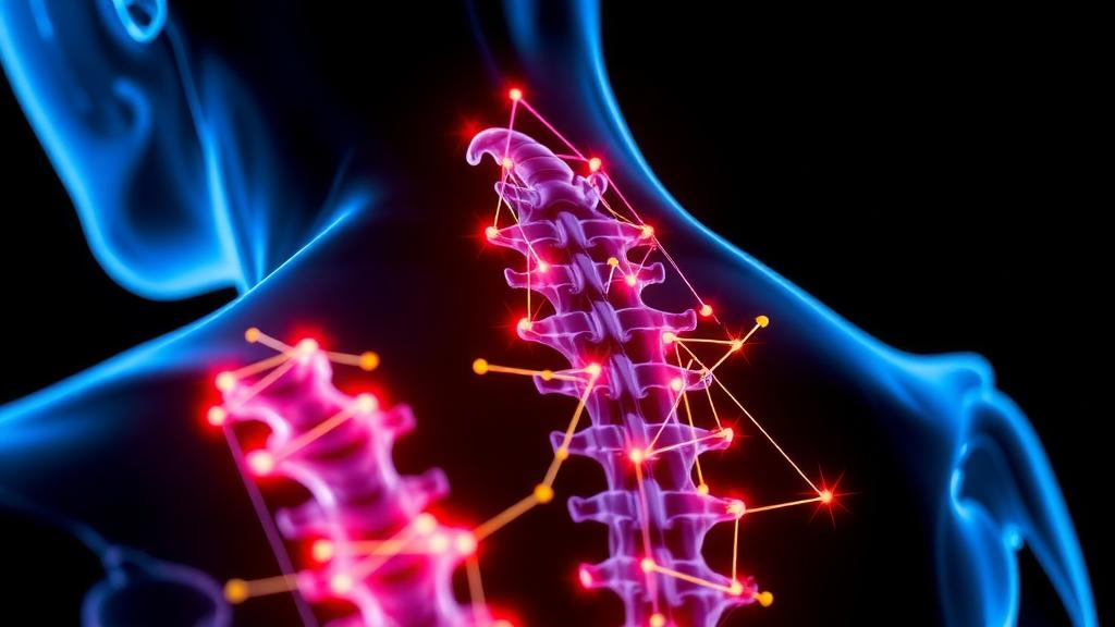
Spinal cord injury remains one of the most devastating conditions in medicine, and one of the hardest to treat. Once the cord is crushed or severed, a cascade of secondary damage unfolds within hours: reactive oxygen species flood the lesion, iron spills from ruptured cells, and neurons die in droves through a specialized form of cell death called ferroptosis. Current clinical care can stabilize patients and ease symptoms, but it cannot reverse the loss of neurons, the destruction of myelin, or the collapse of electrical signaling that leaves tissue below the injury silent. Now, a team of researchers in China has reported a strategy that attacks this cascade on several fronts at once, combining an injectable, antioxidant, electrically conductive hydrogel with stem cell delivery and electroacupuncture, a modernized form of traditional acupuncture that applies microcurrents through needles. The work, published in the journal Materials Today Bio, describes how the combined therapy promoted spinal cord regeneration and restored motor function in injured mice.
The study, led by Yihui Zhang, Yujun Mo, and colleagues including YunGang Wu and Zhouguang Wang, was built around a clear-eyed assessment of why existing approaches fall short. Electroacupuncture is already used clinically in spinal cord injury rehabilitation and can dampen inflammation and boost neurotrophic factors, the signaling molecules that nourish neurons. But on its own, it cannot reverse the death of neurons or repair broken neural circuits, and its effectiveness is limited by the fact that injured tissue conducts electricity poorly. Conductive hydrogels and electroactive scaffolds can partially restore electrical continuity across a lesion, yet they do little to suppress the oxidative storm raging within it. Antioxidant hydrogels, conversely, can mop up reactive oxygen species but lack sustained electrical coupling. And stem cell transplants, however promising, face a brutal reality: bone marrow mesenchymal stem cells, or BMSCs, transplanted into the injury site are themselves vulnerable to the very oxidative stress and ferroptosis that make the environment hostile in the first place.
The researchers’ solution was a composite material they call HBP@BMSCs. The hydrogel matrix is made from dopamine-grafted hyaluronic acid, a polymer that combines the biocompatibility of hyaluronic acid, a natural component of tissue, with the adhesive properties of catechol groups, allowing it to cling tightly to the irregular surfaces of a spinal cord lesion. Suspended within this matrix are nanoparticles of polypyrrole, a well-known conductive polymer, loaded with baicalin, an antioxidant compound derived from the medicinal herb Scutellaria baicalensis. Baicalin does double duty: it directly scavenges free radicals and activates the Nrf2/ARE antioxidant signaling pathway, upregulating endogenous protective proteins such as HO-1, NQO1, and GPX4. The phenolic hydroxyl groups on baicalin also crosslink with polypyrrole to form a conductive nanonetwork whose electrical properties fall within the range of neural tissue, allowing it to carry bioelectric signals through the lesion.
The material itself was carefully engineered. Nuclear magnetic resonance confirmed the dopamine grafting onto hyaluronic acid, and ultraviolet-visible spectroscopy verified that baicalin had been successfully incorporated into the polypyrrole nanoparticles, which grew from roughly 168 to 228 nanometers in diameter after loading. The finished hydrogel gels in about five minutes at body temperature, can be extruded through a syringe, and exhibits shear-thinning behavior, meaning it becomes less viscous under pressure and can flow into the complex geometry of a wound cavity before re-solidifying. At an optimized baicalin-polypyrrole concentration of 0.6 milligrams per milliliter, the hydrogel reached a conductivity of approximately 6 to 7 millisiemens per centimeter, comparable to other conductive hydrogels used in spinal cord repair, while keeping cell viability above 98 percent. Baicalin was released gradually from the matrix over roughly 240 hours, providing sustained antioxidant protection during the most dangerous window of secondary injury.
In laboratory tests, the hydrogel proved remarkably protective. When BMSCs or PC12 cells, a neuron-like model line, were exposed to tert-butyl hydroperoxide to induce oxidative stress, or to erastin, a chemical that triggers ferroptosis, the presence of the HBP hydrogel dramatically reduced reactive oxygen species accumulation and reversed the rise of 4HNE, a hallmark marker of lipid peroxidation, along with ferritin light and heavy chains that signal iron overload. Flow cytometry confirmed that BMSCs cultured inside the hydrogel for 24 hours retained their stem cell identity, with more than 95 percent expressing the mesenchymal markers CD73 and CD90 and fewer than 2 percent expressing the hematopoietic markers CD34 and CD45. In other words, the material sheltered the cells without changing what they were.
The in vivo experiments were conducted in adult female C57BL/6 mice subjected to a standardized compression injury at the T9-T10 vertebral level, with seven treatment groups ranging from saline controls to the full combination of hydrogel-delivered BMSCs plus electroacupuncture. Investigators assessing behavior and quantifying images were blinded to group allocation. Electroacupuncture was administered twice daily for 14 consecutive days at the GV14 and GV4 acupoints, using a 1.0 milliamp current at 4/10 hertz for 15 minutes per session. Seven days after injury, the combined therapy group showed the lowest levels of 4HNE and the highest levels of GPX4, the glutathione-dependent enzyme that stands as the principal brake on ferroptosis. Western blotting revealed that the combination suppressed the oxidative stress proteins COX2 and NOX2 while restoring XCT, GPX4, and SOD1, and that it most strongly activated Nrf2, the master transcriptional regulator of antioxidant defense.
The therapy also tackled the iron problem head-on. After spinal cord injury, disruption of the blood-spinal cord barrier and hemorrhage cause iron to accumulate abnormally in the lesion. Transferrin receptor, which imports iron, rises while ferroportin, which exports it, falls, and ferritin stores swell as cells struggle to buffer the excess. Prussian blue staining showed abundant iron deposits in untreated injured mice, and transcriptome sequencing confirmed that gene sets for iron ion transport and intracellular iron homeostasis were preferentially enriched toward the injury phenotype. The HBP@BMSCs plus electroacupuncture group reversed this pattern most decisively, reducing ferritin and transferrin receptor expression while restoring ferroportin, thereby relieving the intracellular iron load that fuels the Fenton reaction and the vicious cycle of lipid peroxidation. Neuronal apoptosis, measured by cleaved caspase-3 and the Bax-to-Bcl-2 ratio, fell in parallel, and more neurons survived in the lesion core.
Perhaps the most striking results concerned regeneration. Thirty days after treatment, CM-DiI-labeled transplanted BMSCs in the combination group showed markedly increased colocalization with NeuN, a mature neuronal marker, indicating that the cells had differentiated toward a neural lineage. Corticospinal tract tracing, performed by injecting a light-sensitive mCherry-expressing virus into the sensorimotor cortex, revealed abundant regenerating fibers extending through and beyond the lesion in the combination group, whereas injured controls showed almost none. NF200-positive neurofilaments crossed the glial scar boundary marked by GFAP, and the scarred area itself shrank. The conductive BA-PPy component proved essential: a control group receiving BMSCs in plain hyaluronic acid hydrogel with electroacupuncture, but without the nanoparticles, showed substantially weaker neurotrophic signaling, less neuronal differentiation, and poorer circuit reconstruction, demonstrating that the material was actively conducting and amplifying the electrical stimulation rather than merely serving as a passive carrier.
Function followed structure. Using the Basso Mouse Scale and an AI-powered gait analysis system based on DeepLabCut pose tracking, the researchers quantified hindlimb recovery over 30 days. Mice receiving the combined therapy achieved the highest BMS scores, greater muscle strength, longer gait cycles, and higher hip and toe heights, with joint movement amplitudes at the knee, ankle, and toe significantly improved compared with every other group. Motor-evoked potentials recorded from the gastrocnemius muscle after stimulation above the lesion reached their greatest amplitude in the combination group, confirming that signals could once again traverse the injury site. Histology showed preserved tissue architecture, reduced cavity formation, and abundant Nissl bodies, the protein-synthesis machinery of healthy neurons, in the lesion area. Major organs examined 30 days after treatment showed no pathological abnormalities, suggesting the treatment was systemically safe.
The study stops short of human application, and the hurdles between a mouse model and a clinical therapy for spinal cord injury are formidable. But the conceptual advance is significant: rather than treating the biochemical and bioelectrical dimensions of spinal cord injury separately, the researchers fused them into a single intervention in which a conductive, antioxidant, cell-laden matrix transforms electroacupuncture from a modest symptomatic therapy into a driver of genuine regeneration. By simultaneously scavenging reactive oxygen species, restoring iron homeostasis, blocking ferroptosis, protecting transplanted stem cells, and transmitting electrical cues that steer those cells toward neural fates, the platform addresses the interlocking failures that have limited regenerative medicine after spinal cord trauma. If the synergy holds in larger animal models, the humble needle, paired with an engineered gel, may yet become an unexpected centerpiece of spinal cord repair.
Subject of Research: A conductive anti-ferroptosis hydrogel combined with electroacupuncture for spinal cord injury regeneration
Article Title: Electroacupuncture synergizes with anti-ferroptosis bioelectrical niche remodeling to promote spinal cord regeneration
Article References: Zhang, Y., Mo, Y., Yan, B., Wang, X., Wu, P., Wu, Y., & Wang, Z. (2026). Electroacupuncture synergizes with anti-ferroptosis bioelectrical niche remodeling to promote spinal cord regeneration. Materials Today Bio, 41, Article 103708. https://doi.org/10.1016/j.mtbio.2026.103708
Image Credits: AI Generated
DOI: 10.1016/j.mtbio.2026.103708
Keywords: spinal cord injury, electroacupuncture, ferroptosis, hydrogel, stem cells, polypyrrole, baicalin, oxidative stress, iron homeostasis, neural regeneration, biomaterials, conductive scaffold
Cite Scienmag News
Denise Maddox. (October 3, 2026). Electroacupuncture Meets a Smart Hydrogel to Rebuild Injured Spinal Cords. Scienmag. https://scienmag.com/electroacupuncture-meets-a-smart-hydrogel-to-rebuild-injured-spinal-cords/
Denise Maddox. "Electroacupuncture Meets a Smart Hydrogel to Rebuild Injured Spinal Cords." Scienmag, 3 October 2026, https://scienmag.com/electroacupuncture-meets-a-smart-hydrogel-to-rebuild-injured-spinal-cords/. Accessed 4 October 2026.
Denise Maddox. "Electroacupuncture Meets a Smart Hydrogel to Rebuild Injured Spinal Cords." Scienmag. October 3, 2026. https://scienmag.com/electroacupuncture-meets-a-smart-hydrogel-to-rebuild-injured-spinal-cords/


