


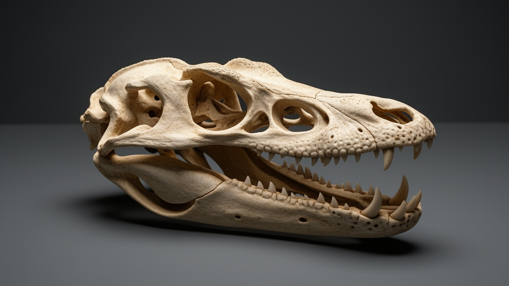
The Komodo dragon has long captured the imagination of scientists and the public alike, but its skull has never been examined quite like this. In a study published in Evolutionary Systematics, researchers used high-resolution CT scanning and 3D digital reconstruction to perform a complete digital dissection of the skull of Varanus komodoensis, the largest living monitor lizard. The result is a detailed three-dimensional map of the cranial skeleton, revealing how the anatomy of this formidable predator is finely tuned for its distinctive feeding behavior and shedding new light on its evolutionary relationships.
The specimen at the heart of the study came from the Zoological Museum Hamburg, cataloged under ZMH R13322. Originally from a captive breeding program at the Washington Zoo, the female Komodo dragon was transferred to the Berlin Zoological Garden in October 1995, weighing 2.56 kg and measuring 750 mm at the time. She died in July 2001 and was preserved frozen at the Hamburg museum for over two decades. By the start of the investigation, her total body length had reached 2030 mm, though prolonged frozen storage had induced a freeze-drying effect that stiffened the specimen and potentially caused some shrinkage.
The CT scan was conducted at Comet Yxlon GmbH in Hamburg using a high-resolution FF85 CT scanner equipped with a 450 kV mini-focus X-ray tube and a Varex Imaging 4343HE detector. The scan produced 3420 cross-sectional images, each sized at 9.4 MB, with 2768 images of the skull used for segmentation, resulting in an initial dataset of 26.02 GB. The cone beam CT settings included a stop-and-go scan type, an X-ray tube power of 570 W operating at 300 kV and 1900 μA, with no filter. Each of the 3052-by-3052-pixel projections had an exposure time of 400 ms, and the total scan duration was just under two hours. The setup yielded a voxel size of 0.0926 mm in each dimension, allowing all measurements to be calculated from voxel size.
Segmentation was performed using AMIRA 6.2.0 software on a workstation with 128 GB RAM. Each bone was independently segmented, primarily using the Magic Wand tool, a region-growing algorithm that selects contiguous voxels within a specified gray-value tolerance. When bones closely neighbored each other with similar gray values, the Draw limit line function allowed manual separation of adjacent elements. After segmentation, the Generate Surface function extracted polygon surfaces from the results, and the Simplification Editor reduced excessive triangle counts. Despite the substantial RAM available, the handling of such large datasets occasionally caused software crashes, and the fine serrations of the teeth remained challenging to render in the 3D model due to resolution limitations.
The reconstructed skull measures 16.72 cm in length, 8.3 cm in width, and 6.46 cm in height, approximating a blunt pyramidal shape with triangular contours in all views, tapering anteriorly. The skull is strongly built but features several large windows, especially in the narial region and the posterior part. Almost completely covered by osteoderm, the skull displays diverse platelet shapes: large flattened rosette-like structures on the dorsal rostrum, simpler plate-like elements on the posterior nasal, rosette-shaped, plate-shaped, and dendritic forms in the occipital region, and long narrow vermiform structures arranged obliquely laterally. Notably, the osteoderm is reduced or absent in the parietal area, at the frontoparietal suture, at the parietal foramen, along the snout, labial margins, and ears, and it makes no contact with the underlying cranial bones.
The digital dissection revealed a total of 30 paired and unpaired bones, each carefully reconstructed. The premaxillae are fused into a single element bearing eight teeth, while each maxilla accommodates thirteen tooth positions with sharp, laterally flattened, posteriorly curved teeth, each accompanied by three to four smaller replacement teeth. The dentary similarly bears twelve teeth per side. The teeth are classified as ziphodont, pleurodont, and polyphodont, reflecting continuous tooth replacement and a feeding strategy optimized for slicing through flesh. The septomaxilla covers Jacobson’s organ, the vomer features deep grooves and ridges, and the palate includes paired palatines, ectopterygoids, and pterygoids with complex processes. The braincase comprises the supraoccipital, prootics, basisphenoid with its fused parasphenoid, paired otooccipitals, and the basioccipital, each contributing to a highly fenestrated posterior skull.
The mandible is equally detailed. The dentary forms the most anterior part, with a long Meckelian groove on its medial side mostly covered by the splenial. The coronoid has three processes, the surangular constitutes roughly half the mandible length, and the articular forms the posteriormost element, receiving the mandibular condyle of the quadrate at the articular fovea. The angular is slender, and the splenial features three major pointed processes. The lower temporal bar is absent, a reduction linked to the evolution of streptostyly, enabling the quadrate to oscillate freely in anterior and posterior directions. This mobile quadrate, combined with loose articulations in the mandible, allows monitor lizards to ingest large food items beyond the capacity of other lizards.
The study identified several anatomical indicators of cranial kinesis in the Komodo dragon skull. A postorbital bar is present, but the orbital process of the postorbitofrontal is not connected to the temporal process of the jugal, providing evidence of mesokinesis. An intracranial joint between the frontal and parietal was observed, a hallmark of mesokinetic skulls. The absence of osteoderm at the frontoparietal suture, functioning as an intracranial joint, may be considered a morphological correlate of skull mesokinesis. Loose connections between various cranial segments, including between the supraoccipital and parietal, between the prootic and parietal, and between the basipterygoid process and pterygoid, were interpreted as mobile connections possibly facilitating the swallowing of large prey objects.
The cranial morphology of the Komodo dragon exhibits pronounced adaptation to its distinctive hold-and-pull feeding technique. The highly fenestrated skull, characterized by a space-frame design, is considered optimized for concurrently executing a jaw adductor-driven bite and a postcranially generated pull-back. The ziphodont teeth, coupled with caudal head movements, facilitate deep wounds in prey and tear flesh into manageable pieces. Intriguingly, the Komodo dragon exhibits a significantly lower bite force compared to other vertebrates of similar body mass, but this is complemented by a strong pulling force in the ventrocaudal direction that exceeds the bite itself. The species also employs coagulopathic, hypotensive, hemorrhagic, and shock-inducing toxins during prey capture, with venom glands positioned between the teeth.
Beyond feeding adaptations, the study evaluated the phylogenetic position of the Komodo dragon. All eleven cranial synapomorphies previously identified for Saniwa plus Varanus were validated in the skull, and comparison with macerated skulls of four other varanid species confirmed these shared derived characters. Furthermore, six unequivocal synapomorphies within the braincase were identified as shared between the Komodo dragon and the extinct giant monitor Varanus priscus, known as Megalania, supporting a close relationship between the two species. These shared features, combined with the space-frame skull design and ziphodont tooth morphology reminiscent of theropod dinosaurs like Allosaurus, suggest convergent evolution of biomechanical adaptations for large-prey predation across distantly related predators, offering a vivid example of how skeletal form mirrors function in the natural world.
Subject of Research: The skull morphology of the Komodo dragon, Varanus komodoensis (Reptilia, Squamata, Varanidae) — a digital-dissection study
Article Title: The skull morphology of the Komodo dragon, Varanus komodoensis ( Reptilia , Squamata , Varanidae ) — a digital-dissection study
Article References: Kubota, M., Hallermann, J., Beerlink, A., & Haas, A. (2024). The skull morphology of the Komodo dragon, Varanus komodoensis (Reptilia, Squamata, Varanidae) — a digital-dissection study. Evolutionary Systematics, 8(2), 219-245. https://doi.org/10.3897/evolsyst.8.121149
Image Credits: AI Generated
DOI: 10.3897/evolsyst.8.121149
Keywords: skull, morphology, Komodo, dragon, Varanus, komodoensis, Reptilia, Squamata, Varanidae, digital-dissection, scientific research
Cite Scienmag News
Drew Townsend. (October 19, 2026). Digital dissection reveals how the Komodo dragon skull is built for hold-and-pull predation. Scienmag. https://scienmag.com/digital-dissection-reveals-how-the-komodo-dragon-skull-is-built-for-hold-and-pull-predation/
Drew Townsend. "Digital dissection reveals how the Komodo dragon skull is built for hold-and-pull predation." Scienmag, 19 October 2026, https://scienmag.com/digital-dissection-reveals-how-the-komodo-dragon-skull-is-built-for-hold-and-pull-predation/. Accessed 19 October 2026.
Drew Townsend. "Digital dissection reveals how the Komodo dragon skull is built for hold-and-pull predation." Scienmag. October 19, 2026. https://scienmag.com/digital-dissection-reveals-how-the-komodo-dragon-skull-is-built-for-hold-and-pull-predation/


