


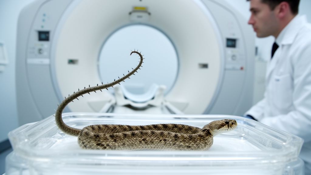
X-ray computed tomography has revealed that the conspicuous, spider-shaped tail tip of the Iranian spider-tailed horned viper is supported by vertebrae that are typical of most vipers. Researchers studying the preserved holotype of Pseudocerastes urarachnoides found no comparably unusual internal bony structure corresponding to the bulbous, leg-like formation on the outside of the tail. The study provides the first internal view of the animal’s celebrated bird-luring appendage and shows that its extraordinary appearance is not matched by extraordinary terminal anatomy. Published in The Anatomical Record, the anatomical study concerns one preserved specimen and its vertebral column; it does not directly test how the tail develops or how birds respond to it.
The snake examined was the species’ holotype, the specimen formally used as the standard reference for its physical characteristics. It was collected in 1968 and taken to the Field Museum in Chicago, where staff initially interpreted the tip of its tail as a deformity resembling a tumor. The specimen remained in the collection for decades. Its unusual appearance prompted renewed attention when another snake with a similar tail was found in 2003, and scientists formally described Pseudocerastes urarachnoides in 2006 using the Field Museum specimen as the holotype. The new work did not newly discover the species or the bird-luring behavior; it applied a newer imaging method to a long-known animal to examine the anatomy behind its visible tail.
Field observations had previously documented how the viper uses the appendage at the end of its tail. Its tip resembles a small spider, with a bulbous body and long, dangling legs, and the snake twitches this structure as birds approach. Video recorded in the wild showed birds swooping toward the moving spider-like form, after which the snake could strike and consume a bird. The behavior is a specialized form of mimicry in which the tail resembles an animal capable of attracting prey. Those observations supplied the behavioral context for the anatomical study, but they were separate from the XCT examination. The scans were performed to determine what lay beneath the unusual exterior, rather than to establish whether the tail could function as a lure.
Snake vertebrae can differ markedly from one position along the body to another, making isolated fossil bones difficult things to identify with confidence. The researchers were particularly interested in whether the spider-tailed horned viper had terminal vertebrae as strange in form as some of those found in sand boas and rubber boas. Those snakes can possess conspicuously shaped bones at the ends of their tails, providing a relevant comparison for a viper whose external tail tip is so unusual. The anatomical question was therefore specific: did the spider-like appendage contain a similarly alien skeletal architecture? The study did not seek a broad survey of viper skeletons, nor did it compare numerous individuals of the species.
To examine the holotype without cutting into it, the team used X-ray computed tomography, also referred to as XCT. The method produces thousands of X-ray images and combines them into a detailed three-dimensional rendering of structures inside an object. Unlike physical dissection, it allows researchers to inspect internal anatomy without removing or damaging the specimen. That distinction was particularly important for a taxonomically important snake, because the holotype serves as the physical reference against which characteristics of the species are compared. The Field Museum’s new XCT laboratory began operating in late 2024, and the spider-tailed horned viper was among the first specimens scanned after it opened.
The resulting three-dimensional images showed a tail skeleton that looked broadly normal for a viper. According to study first author Georgios Georgalis of the Polish Academy of Sciences in Kraków, the spider tip is external morphology: the tail vertebrae themselves were typical of most vipers, and nothing in the skeleton suggested how extreme the tail looks from the outside. The scan did not expose a hidden specialized structure behind the spider-like form. Instead, the central anatomical finding is a mismatch between outward appearance and underlying vertebral form. The study provides no evidence that the vertebrae produce the lure or determine its movement, and it does not identify the tissue-level mechanism responsible for creating the bulbous body and leg-like projections.
The result offers a modern example of the interpretive limits faced by paleontologists. Fossil snakes are frequently represented by individual vertebrae rather than complete skeletons, and viper vertebrae can provide few clues about the identity of an animal when no other anatomy is preserved. For Georgalis, a paleontologist, this makes the spider-tailed horned viper a useful comparison because its skeletal material does not advertise its most conspicuous feature or the predatory behavior associated with that feature. A researcher working only from isolated bones would have little basis for imagining the extraordinary tail tip. The case illustrates that preserved skeletons can constrain scientific interpretation while omitting features that shape an animal’s appearance and ecological role.
The finding also creates a foundation for questions about how the external tissues of the tail evolve and develop, while leaving those questions unresolved. The XCT analysis establishes that the conspicuous appendage is not accompanied by comparably unusual terminal vertebrae. It does not, however, describe the tissue composition of the spider-like tip, trace its growth, or demonstrate the developmental processes that produce its shape. The authors’ broader significance lies in showing that skeletal form alone is insufficient for inferring every aspect of an animal’s biology. A future investigation of the appendage’s development would need evidence beyond the vertebral patterns examined here, rather than treating the present scan as a complete account of its construction.
For co-author Sara Ruane, the project demonstrates that museum collections can remain scientifically productive long after specimens are collected. The 1968 snake helped establish the species when it was described in 2006 and yielded its internal tail anatomy more than five decades later using technology unavailable at the time of collection. The specimen also illustrates how an object initially interpreted as a deformity can become central to new research once comparable animals are found and its unusual function is documented. XCT did not reveal a concealed mechanism that made the bird-luring behavior work. It revealed a striking contrast instead: the snake’s skeletal tail is comparatively ordinary, while the external structure that attracts birds is anything but.
Subject of Research: Comparative vertebral anatomy of the spider-tailed horned viper’s external bird-luring tail tip
Article Title: A fake spider at the tip of this snake’s tail helps it lure birds. Scientists got their first look inside the “spider.”
Article References: A fake spider at the tip of this snake’s tail helps it lure birds. Scientists got their first look inside the “spider.”. Original publication (Bibliographic details unavailable.)
Image Credits: AI Generated
DOI: Not provided
Keywords: spider-tailed horned viper, Pseudocerastes urarachnoides, X-ray computed tomography, snake anatomy, vertebral morphology, bird-luring mimicry, museum collections, paleontology, Iranian viper, fake, spider, snake
Cite Scienmag News
Drew Townsend. (October 26, 2026). CT scan finds spider-tailed viper’s bird-luring tail has typical vertebrae. Scienmag. https://scienmag.com/ct-scan-finds-spider-tailed-vipers-bird-luring-tail-has-typical-vertebrae/
Drew Townsend. "CT scan finds spider-tailed viper’s bird-luring tail has typical vertebrae." Scienmag, 26 October 2026, https://scienmag.com/ct-scan-finds-spider-tailed-vipers-bird-luring-tail-has-typical-vertebrae/. Accessed 26 October 2026.
Drew Townsend. "CT scan finds spider-tailed viper’s bird-luring tail has typical vertebrae." Scienmag. October 26, 2026. https://scienmag.com/ct-scan-finds-spider-tailed-vipers-bird-luring-tail-has-typical-vertebrae/


