

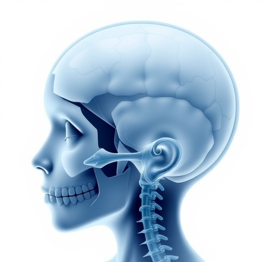
For decades, craniosynostosis has been viewed primarily as a problem of carpentry: an infant’s skull fuses too early, and the surgeon’s job is to remodel the misshapen vault to give the brain room to grow. Yet a landmark review published this week in Pediatric Research argues that this perspective is dangerously narrow. The premature fusion of the cranial sutures, the fibrous joints that normally remain pliable throughout early childhood, may not be merely a mechanical issue but an early warning signal of a far broader disruption to brain development. The paper, led by researchers Vieira, Fleischman Henrique, and de Paula Pereira Henrique, reframes craniosynostosis as a global neurodevelopmental risk, not just a surgical condition, and calls for a radical rethinking of how these children are monitored and supported long after the operating room lights have dimmed.
Craniosynostosis occurs in roughly one in 2,000 live births and has long been classified into syndromic forms, such as Apert or Crouzon syndromes, where mutations in genes like FGFR2 or TWIST1 are well-established, and nonsyndromic cases that appear in isolation. The majority of infants fall into the latter camp. For them, the standard of care is a strip craniectomy or more extensive cranial vault reconstruction ideally performed within the first year of life. The goal has always been twofold: to restore a normal head shape and, crucially, to relieve any potential elevation of intracranial pressure that could, if unaddressed, impair brain growth and lead to cognitive deficits. In the collective surgical imagination, fixing the skull effectively fixed the problem.
But a growing body of literature, meticulously synthesized by Vieira and colleagues, suggests that this reassuring narrative crumbles upon closer inspection. The team meticulously combed through longitudinal neurodevelopmental outcomes, neuroimaging studies, and molecular analyses to paint a picture that is both more subtle and more alarming. They found that even after surgically successful repairs, a significant subset of children with nonsyndromic craniosynostosis continue to demonstrate delays in language, motor skills, attention, and executive function compared to their peers. These deficits often emerge not in the immediate postoperative period but during the critical synaptic pruning and rewiring phases of late toddlerhood and early school age—a timeline that points to a fundamental, underlying dysregulation of neural circuitry rather than a transient effect of elevated pressure.
The technical explanation delves into the shared embryological origin of the cranial sutures and the meninges, the protective membranes encasing the brain. The dura mater, the outermost of these layers, serves as a critical signaling center, secreting a cocktail of growth factors, including fibroblast growth factors and bone morphogenetic proteins, that guide both suture patency and cortical development. When the molecular machinery goes awry—whether due to a single gene mutation or more complex polygenic and environmental interactions—the fusion of a suture may be the most visible manifestation of a process that also subtly alters cortical lamination, interneuron migration, and the formation of thalamocortical connections. In other words, the skull is not simply constricting a healthy brain; the brain itself may be wired differently from the start. Advanced diffusion tensor imaging studies cited in the review reveal reduced white matter integrity and atypical connectivity patterns in children with single-suture fusions, abnormalities that often persist regardless of whether the suture was surgically released.
This reframing has immediate, actionable consequences. If craniosynostosis is a skeletal red flag for atypical neurodevelopment, then surgical correction, while still essential to prevent the most obvious physical complications, cannot be the endpoint of care. The authors argue for a standardized, multidisciplinary surveillance protocol that follows affected children at least through the early school years, incorporating regular neuropsychological assessments and early intervention therapies that target the specific cognitive domains most at risk, such as working memory and visual-motor integration. Currently, many children with isolated sagittal synostosis, the most common form, are discharged from neurosurgical follow-up with a clean bill of health once the shape is normalized, leaving families unprepared for the learning struggles that may surface later.
One of the most provocative ideas outlined in the paper is the possibility that the neurodevelopmental risk exists on a spectrum, tied to the specific suture that fuses. For example, metopic synostosis, which creates a triangular forehead, appears to carry a higher burden of behavioral and attention difficulties, potentially due to the impact on frontal lobe development pathways. In contrast, sagittal synostosis is more frequently associated with language delays. This pattern specificity further undermines a one-size-fits-all mechanical model and reinforces the hypothesis that local suture biology and regional brain development are intimately linked, with each suture acting as a sentinel for the underlying cortical territory.
The team also highlights how the reclassification of craniosynostosis as a global developmental risk could unlock new therapeutic avenues. If key molecular pathways like the FGF signaling cascade are misfiring in both the suture mesenchyme and the developing forebrain, agents that modulate these pathways—some already in preclinical testing for related skeletal dysplasias—might one day be deployed not just to prevent suture fusion in utero but to shepherd more typical brain development. Such pharmacological strategies remain speculative, but they mark a profound conceptual shift from a purely mechanical fix to a neuroprotective strategy that targets the biology at its root.
The study does not diminish the life-changing importance of cranial surgery, which remains a brilliant and necessary technical achievement. Instead, it demands that the medical community see beyond the millimeter-thin suture line and recognize craniosynostosis for what it truly is: an integrated disorder of skull and brain development that requires integrated, long-term care. As one of the authors summarized in an accompanying statement, “We have been treating the scaffold while overlooking the organ it houses. The evidence now tells us we must do both.”
Subject of Research: The relationship between craniosynostosis and long-term neurodevelopmental outcomes, reframing the condition from a purely surgical issue to a global developmental risk.
Article Title: Craniosynostosis and neurodevelopment: reframing a surgical condition as a global developmental risk
Article References: Vieira, B.A., Fleischman Henrique, C.C., de Paula Pereira Henrique, R. et al. Craniosynostosis and neurodevelopment: reframing a surgical condition as a global developmental risk. Pediatr Res (2026). https://doi.org/10.1038/s41390-026-05249-0
Image Credits: AI Generated
DOI: 10.1038/s41390-026-05249-0
Keywords: Craniosynostosis, neurodevelopment, pediatric neurosurgery, developmental risk, skull sutures, brain development, surgical conditions, genetic syndromes, FGFR2, TWIST1, dural signaling, white matter integrity, diffusion tensor imaging, neuropsychological outcomes, early intervention.


