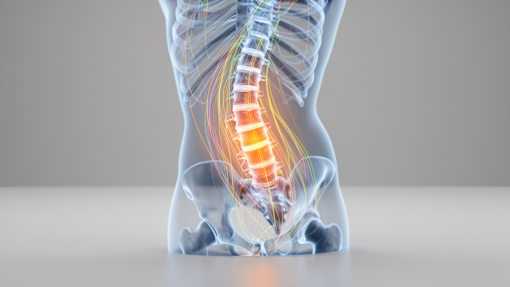
Corrective osteotomy for severe spinal deformity requires shortening the vertebral column, yet surgeons have lacked a clear biomechanical answer to a deceptively simple question: how much shortening is too much? A new modeling study published in Scientific Reports addresses that question using fluid-structure interaction finite element analysis, simulating how graded vertebral shortening alters cerebrospinal fluid dynamics and perfusion of the anterior spinal artery.
The research team, led by Alimujiang Yusufu and colleagues, reconstructed a three-dimensional model of the thoracic spine from T1 to T12 using computed tomography data obtained from a 16-year-old male patient with scoliosis. Because the model was built from a single patient’s imaging, the findings are explicitly patient-specific rather than generalizable to all spinal geometries.
Within this model, the researchers simulated posterior vertebral column resection at the T8/T9 level under four conditions: no shortening at all, and shortening by one-third, one-half, and two-thirds of a vertebral segment. The simulations coupled a cerebrospinal fluid-dura model with a blood-vessel wall model, allowing the team to quantify pressure, wall shear stress, flow velocity, structural stress, and displacement across both the fluid and solid compartments.
The results revealed a strikingly stable regime at moderate shortening. When the column was shortened by up to one-half, cerebrospinal fluid wall shear stress held at approximately 0.04 pascals and fluid velocity at approximately 0.014 meters per second, values that remained essentially unchanged across that range.
At the same time, the anterior spinal artery showed a different pattern. Luminal pressure and blood flow velocity declined gradually as shortening increased, together with vessel-wall stress and displacement. According to the authors, this combination indicates preserved hemodynamic compensation alongside what they describe as mechanical unloading of the vessel, meaning the artery experienced progressively less mechanical load even as its perfusion variables stayed within a compensated range.
The picture changed dramatically at two-thirds shortening. Cerebrospinal fluid wall shear stress rose to 0.4998 pascals, an increase of roughly twelve-fold, while fluid velocity climbed to 0.16009 meters per second, about eleven-fold higher. Dural stress increased by approximately 34 percent, yet displacement rose by only 1.2 percent, a disparity the authors describe as nonlinear stress-displacement decoupling.
That decoupling is technically significant because it shows that structural deformation alone would not have signaled the danger. The dura barely moved while the stresses acting upon it and the fluid dynamics within it changed by an order of magnitude, meaning displacement-based assessments could substantially underestimate the mechanical burden imposed by aggressive shortening.
The anterior spinal artery variables also rebounded at the two-thirds level, with luminal pressure, velocity, vessel-wall stress, and displacement all reaching their highest simulated values. In the model’s terms, the vascular unloading seen at moderate shortening reversed, and the artery returned to its most mechanically loaded state of any condition tested.
Taken together, the authors conclude that in this patient-specific model, shortening up to one-half was associated with relative stability of cerebrospinal fluid dynamics and progressive vascular unloading, whereas two-thirds shortening produced marked nonlinear changes in both cerebrospinal fluid and anterior spinal artery biomechanical variables. They characterize this as a model-derived biomechanical transition located between one-half and two-thirds shortening.
The team is careful to bound the clinical meaning of the result. The transition point emerges from a single computational model of one patient’s spine, and the authors state explicitly that it should not be interpreted as a clinically validated safety threshold.
Subject of Research: Fluid-structure interaction modeling of vertebral shortening during corrective osteotomy for spinal deformity
Article Title: Effects of vertebral shortening during corrective osteotomy for spinal deformity on spinal cord biomechanics and cerebrospinal fluid-anterior spinal artery fluid-structure interaction
Article References: Yusufu, A., Abulaiti, A., Haibier, A., & Ma, Y. (2026). Effects of vertebral shortening during corrective osteotomy for spinal deformity on spinal cord biomechanics and cerebrospinal fluid-anterior spinal artery fluid-structure interaction. Scientific Reports. https://doi.org/10.1038/s41598-026-71867-7
Image Credits: AI Generated
DOI: 10.1038/s41598-026-71867-7
Keywords: spinal deformity, corrective osteotomy, vertebral shortening, cerebrospinal fluid, anterior spinal artery, fluid-structure interaction, finite element analysis, spinal cord biomechanics, posterior vertebral column resection, scoliosis, wall shear stress, biomechanical modeling
Cite Scienmag News
Ophelia Keating. (October 29, 2026). Computer model maps a biomechanical tipping point in spinal deformity surgery. Scienmag. https://scienmag.com/computer-model-maps-a-biomechanical-tipping-point-in-spinal-deformity-surgery/
Ophelia Keating. "Computer model maps a biomechanical tipping point in spinal deformity surgery." Scienmag, 29 October 2026, https://scienmag.com/computer-model-maps-a-biomechanical-tipping-point-in-spinal-deformity-surgery/. Accessed 31 October 2026.
Ophelia Keating. "Computer model maps a biomechanical tipping point in spinal deformity surgery." Scienmag. October 29, 2026. https://scienmag.com/computer-model-maps-a-biomechanical-tipping-point-in-spinal-deformity-surgery/