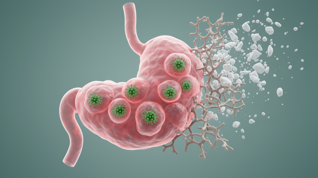
Epstein-Barr virus-associated gastric cancer (EBVaGC) has long been regarded as the most immunologically favorable form of a disease that remains the fifth most common cancer worldwide. Accounting for roughly 4.1 to 9 percent of all gastric cancers, this molecularly distinct subtype carries recurrent PIK3CA mutations, extreme DNA hypermethylation, and amplification of JAK2 and PD-L1/PD-L2, and it presents viral antigens that attract dense infiltrates of CD3-positive T cells and CD68-positive macrophages. Yet a new study published in Nature Communications suggests that the standard treatment for locally advanced disease, neoadjuvant chemotherapy, may inadvertently sabotage the very immune architecture that gives this virus-driven cancer its better prognosis.
The research, led by Jiwang Bi, Shuofeng Hu, Yu Sun, Ting Guo, Jiafu Ji, and Xiaofang Xing, centered on tertiary lymphoid structures (TLS), organized aggregates of immune cells that form within tumors and act as local hubs for anti-cancer immune activation. TLS are strongly correlated with therapeutic efficacy and patient survival across many cancers, and their maturation status critically shapes outcomes: mature structures with well-formed germinal centers predict favorable survival, whereas immature ones predict poor immunotherapy response. How neoadjuvant chemotherapy affects these structures in a highly immune cell-infiltrated, virus-associated microenvironment had remained unexplored.
To address this gap, the team assembled a large single-center cohort of 144 locally advanced EBVaGC patients, comprising 99 who were treatment-naive and 45 who received fluorouracil and platinum-based neoadjuvant chemotherapy, alongside 1,352 EBV-negative gastric cancer cases for comparison. The investigators combined pathological evaluation, multiplex immunohistochemistry, bulk and single-cell RNA sequencing from 77 gastric cancer samples, and spatial transcriptomics from 11 representative EBVaGC specimens. This multi-omics strategy allowed them to map TLS density, maturity, and spatial organization with unprecedented resolution across the tumor microenvironment.
The first key finding confirmed the immunogenic reputation of EBVaGC. Compared with EBV-negative tumors, EBVaGC exhibited significantly greater infiltration of T cells, B cells, dendritic cells, and monocytic lineage cells. At the single-cell level, the proportions of TLS-associated components, including germinal center B cells, activated LAMP3-positive dendritic cells, and follicular helper T cells, were markedly higher. Although quantitative TLS density did not differ significantly between the two subtypes, EBVaGC displayed dramatically increased TLS maturity, with organized structures accounting for 35.1 percent versus 14.6 percent of TLS in EBV-negative disease, and well-formed secondary follicle-like TLS with identifiable germinal centers reaching 8.3 percent versus 2.5 percent.
Spatially, the team distinguished TLS located within 1 millimeter of the tumor boundary, termed invasive margin TLS (IM-TLS), from tumor center TLS (TC-TLS) positioned deeper within the parenchyma. Survival analysis of the EBVaGC cohort revealed that the presence of IM-TLS, rather than TC-TLS, served as a significant prognostic stratification factor: patients with IM-TLS experienced significantly prolonged overall survival and disease-free survival. Multivariate Cox regression confirmed that IM-TLS presence was an independent prognostic factor for disease-free survival, with a hazard ratio of 0.45, underscoring the critical importance of these immune structures at the tumor’s invasive frontier.
The clinical results concerning neoadjuvant chemotherapy were sobering. Only one of 45 patients, or 2.2 percent, achieved a pathological complete response following chemotherapy, considerably lower than the 5.6 to 22.3 percent rates reported in unselected gastric cancer populations. Comparative survival analysis showed no significant improvement in overall or disease-free survival for patients who received chemotherapy compared with those who underwent upfront surgery, and chemotherapy was not an independent factor for reducing recurrence risk. Structurally, chemotherapy-treated tumors showed significantly reduced TLS density and impaired maturation, with well-formed secondary follicle-like TLS almost abolished, falling from 8.3 percent to 0.9 percent.
Spatial transcriptomics refined the pathological definition of the invasive margin. By subdividing the tumor border into 20 concentric layers of 100 micrometers each and applying the BANKSY spatial niche algorithm, the researchers identified a functionally active invasive margin spanning only plus or minus 300 micrometers from the tumor border. Within this narrowed zone, transcriptomic profiling revealed significant upregulation of immune activation genes essential for TLS formation, including CCL19, LTB, and IGHM, alongside robust enrichment of T cell receptor and B cell receptor signaling pathways. An independent multiplex immunohistochemistry cohort of 23 samples confirmed that the absolute peak of TLS counts falls exactly within this window, and the refined cutoff successfully stratified patient prognosis.
Within this critical region, chemotherapy fundamentally altered the immune landscape. Post-treatment samples showed significantly reduced abundance of germinal center B cells and activated dendritic cells, whereas follicular helper T cells and regulatory T cells were markedly increased. Among patients who received chemotherapy, those who developed recurrence showed further reduction in germinal center B cells and increased regulatory T cells, accompanied by significantly higher levels of CXCL13-secreting CD8-positive T cells, indicating a more exhausted microenvironment. Neutrophils emerged as the most dramatically altered population: their abundance negatively correlated with germinal center B cells and activated dendritic cells but positively with regulatory T cells, and multiplex immunohistochemistry revealed that NET-producing neutrophils, co-expressing neutrophil elastase and citrullinated histone H3, densely encased immature TLS in relapsed patients while mature structures remained free of such infiltration.
Mechanistically, the team traced this neutrophil dysfunction to chemotherapy-induced Epstein-Barr virus reactivation. Single-cell subtyping identified an aging neutrophil phenotype that downregulated the CD62L marker SELL and upregulated the homing receptor CXCR4, driven by an IRF7-centric regulatory module that strongly upregulated pattern recognition receptors, antiviral interferon-stimulated genes, and NET-formation mediators. Cell-cell communication analysis predicted strong CXCR4-CXCL12 interactions with stromal cells, which the team validated by showing that stromal cells surrounding IM-TLS are the primary producers of CXCL12. In vitro Transwell chemotaxis assays demonstrated that neutrophils indirectly cocultured with chemotherapy-treated EBV-positive SNU719 cells significantly upregulated surface CXCR4 and migrated robustly toward a CXCL12 gradient, whereas coculture with chemotherapy-treated EBV-negative AGS cells failed to induce either response, confirming the EBV-specific nature of the phenomenon.
The viral mechanism was confirmed at multiple levels. Treatment with 5-fluorouracil potently elicited a lytic reactivation signature in EBV-positive cancer cells, marked by massive upregulation of the immediate-early gene BZLF1 and late lytic genes alongside downregulation of latent genes, and this response was not drug-specific, as oxaliplatin and paclitaxel produced similar effects. In situ multiplexed fluorescence in situ hybridization validated active lytic transcription in post-treatment patient tissues. Neutrophils exposed to reactivated EBV-positive cells upregulated the viral sensors CGAS, AIM2, and TLR7, and protein-protein interaction networks linked this pattern recognition receptor signaling directly to NET central hubs including MPO, ELANE, and PRTN3. Functional Transwell assays demonstrated that NETs released by these aging neutrophils acted as a macroscopic physical barrier preventing CFSE-labeled lymphocytes from migrating toward CXCL13, and critically, DNase I treatment and TLR7 inhibition with hydroxychloroquine partially restored migration, whereas anti-PD-1 blockade failed to do so, demonstrating that physical exclusion rather than checkpoint signaling is the dominant rate-limiting barrier. These cells also lacked ARG1 expression, the classical hallmark of polymorphonuclear myeloid-derived suppressor cells, confirming they represent a distinct, virus-driven cellular state.
Clinically, the team used a large multiplex immunohistochemistry cohort of 216 specimens to spatially colocalize EBV reactivation, NETs, and IM-TLS. In relapsed post-treatment patients, the proportion of BZLF1-positive tumor cells correlated significantly and positively with the density of NET-releasing neutrophils at the invasive margin, while the ratio of mature TLS area to total TLS area correlated negatively with both. Spatial distance analysis showed that BZLF1-positive tumor cells lay significantly closer to NET-releasing neutrophils than BZLF1-negative cells, with the viral load peaking within 0 to 20 micrometers of these neutrophils. The density of NET-releasing neutrophils around early TLS was significantly higher than around mature TLS in post-treatment relapsed EBVaGC, a pattern completely absent in EBV-negative groups.
The authors acknowledge important limitations, including the low incidence of EBVaGC, reliance on single-center data, and the absence of immunocompetent animal models for this cancer, meaning the causal relationship between NET accumulation and TLS maturation arrest remains inferred from correlative spatial associations and in vitro systems. Nevertheless, the findings carry substantial therapeutic implications. Because mature TLS are vital hubs for generating anti-tumor T cell responses and robust predictors of immune checkpoint blockade efficacy, the discovery that standard chemotherapy erects a NET-mediated physical barrier around TLS suggests that simply superimposing immunotherapy onto chemotherapy might not yield optimal synergy. The authors propose that future combination strategies should incorporate microenvironment-modulating agents aimed at dismantling these barriers, such as targeted NET degraders like DNase I, TLR7 or CXCR4 antagonists, or selective viral suppressors, potentially alongside broader checkpoint targets including LAG-3. By preserving TLS structural integrity and facilitating unimpeded immune cell trafficking, such barrier-breaking approaches could restore effective TLS-mediated anti-tumor immunity in this virus-associated cancer subtype.
Subject of Research: Neoadjuvant chemotherapy interferes with tertiary lymphoid structure formation in Epstein-Barr virus-associated gastric cancer
Article Title: Neoadjuvant chemotherapy interferes with tertiary lymphoid structure formation in Epstein-Barr virus-associated gastric cancer
Article References: Bi, J., Hu, S., Sun, Y., Guo, T., Deng, Z., Hu, Y., Zhao, X., Li, C., Wang, X., Li, X., Zhong, F., Ying, X., Ji, J., & Xing, X. (2026). Neoadjuvant chemotherapy interferes with tertiary lymphoid structure formation in Epstein-Barr virus-associated gastric cancer. Nature Communications, 17(1), Article 11060. https://doi.org/10.1038/s41467-026-77546-5
Image Credits: AI Generated
DOI: 10.1038/s41467-026-77546-5
Keywords: Neoadjuvant, chemotherapy, interferes, tertiary, lymphoid, structure, formation, Epstein-Barr, virus-associated, gastric, cancer, scientific research
Cite Scienmag News
Nathaniel Bowman. (October 24, 2026). Chemotherapy disrupts immune structures in Epstein-Barr virus-linked gastric cancer. Scienmag. https://scienmag.com/chemotherapy-disrupts-immune-structures-in-epstein-barr-virus-linked-gastric-cancer/
Nathaniel Bowman. "Chemotherapy disrupts immune structures in Epstein-Barr virus-linked gastric cancer." Scienmag, 24 October 2026, https://scienmag.com/chemotherapy-disrupts-immune-structures-in-epstein-barr-virus-linked-gastric-cancer/. Accessed 25 October 2026.
Nathaniel Bowman. "Chemotherapy disrupts immune structures in Epstein-Barr virus-linked gastric cancer." Scienmag. October 24, 2026. https://scienmag.com/chemotherapy-disrupts-immune-structures-in-epstein-barr-virus-linked-gastric-cancer/