


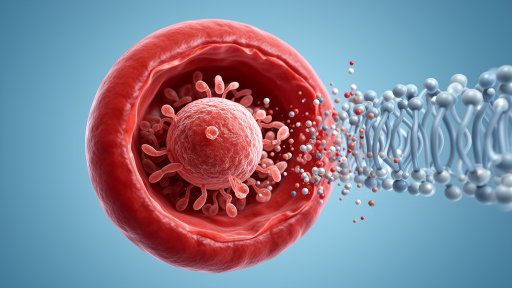
Every 48 hours, Plasmodium falciparum, the parasite responsible for the deadliest form of human malaria, must break out of the red blood cell it has infected to keep its lethal cycle going. This escape, called egress, is not a passive event but a tightly choreographed proteolytic cascade, and researchers at the Francis Crick Institute in London have now rebuilt its final act in a test tube. In a study published in Frontiers in Parasitology, the team describes a cell-free system that reproduces the maturation of SERA6, a parasite cysteine protease-like protein whose activation is the decisive step that allows merozoites to burst free and invade fresh erythrocytes. The work delivers the first direct demonstration that fully processed SERA6 is an enzymatically active protease and validates a new, markedly more potent small-molecule inhibitor, cementing SERA6 as a genuine antimalarial drug target.
The biological context is one of mounting urgency. Malaria still imposes enormous morbidity and mortality worldwide, with children under five and pregnant women at particular risk of severe disease, and chemotherapy remains central to control even as promising vaccines advance. Yet resistance of P. falciparum to artemisinin-based mainstay drugs continues to spread, sharpening the need for target-based drug discovery grounded in a deeper understanding of parasite biology. Egress has emerged as an especially attractive point of intervention because it depends on a defined sequence of parasite enzymes, none of which is targeted by any antimalarial currently in clinical use.
Over the past decade, the egress cascade has been mapped in remarkable detail. It is triggered by activation of a cGMP-regulated parasite kinase called PKG, which itself depends on a guanylyl cyclase, a serine-threonine phosphatase called PP1, the multi-pass membrane protein ICM1 and the calcium-dependent kinase CDPK5. PKG activation discharges a subtilisin-like serine protease, SUB1, from specialized merozoite organelles called exonemes into the lumen of the parasitophorous vacuole that houses the mature schizont. There SUB1 encounters the precursor of SERA6 and cleaves it at two defined sites, releasing a central module called p65, composed of an N-terminal prodomain fused to a papain-like catalytic domain. A rapid autocatalytic step then sheds the prodomain, known as p40, generating the terminal papain-like form, SERA6 PAP.
The final piece of the activation puzzle is a second vacuolar protein, malarial SERA activator 180, or MSA180, which is itself cleaved by SUB1. SERA6 PAP interacts with at least two MSA180 fragments, and the resulting complex is thought to cleave β-spectrin, a core component of the red blood cell cytoskeleton. Destabilization of the cytoskeleton then precipitates rupture of the erythrocyte membrane and the explosive release of merozoites. Consistent with this model, small molecules that block the autocatalytic p65-to-PAP conversion selectively prevent β-spectrin cleavage and red cell rupture while leaving earlier, SERA6-independent events, such as vacuole rounding and rupture and red cell membrane poration, untouched. What remained unclear was whether components beyond SUB1 and MSA180 were needed for maturation, and whether mature SERA6 PAP truly possessed catalytic activity.
Recombinant SUB1 has been available for years, but repeated attempts by the team and others to produce correctly folded recombinant full-length SERA6, full-length MSA180 or SERA6 PAP had failed, stalling mechanistic work. The researchers sidestepped this obstacle by exploiting the parasite itself. Because the non-ionic detergent saponin permeabilizes the red cell and vacuolar membranes without rupturing the parasite plasma membrane, saponin extracts of schizonts are enriched in soluble vacuolar proteins, including full-length SERA6. The team used a transgenic P. falciparum line expressing functional SERA6 with an internal haemagglutinin epitope tag, arrested mature schizonts just before egress with a PKG inhibitor called compound 2, which keeps SUB1 sequestered in exonemes, and prepared saponin extracts predicted to contain everything needed for SERA6 maturation except SUB1 itself.
The prediction held. Adding recombinant SUB1 to these ex vivo extracts triggered the expected time-dependent conversion of full-length SERA6 to PAP via the p65 intermediate, and the epoxide E64, a known inhibitor of the autocatalytic step, blocked the p65-to-PAP conversion. The entire SUB1-dependent maturation pathway had been reconstituted outside the cell, and it remained susceptible to small-molecule inhibition. The team then went further, affinity-purifying tagged full-length SERA6 and MSA180 from the extracts of two transgenic lines and recombining the isolated proteins with recombinant SUB1. Without MSA180, SUB1 converted SERA6 only to the p65 intermediate; with MSA180 present, conversion to PAP was complete. Since both precursors remained stable without added SUB1, the ex vivo reaction faithfully mimicked the in vivo process and showed that SUB1 and MSA180 are both necessary, and apparently sufficient, for full SERA6 maturation, with no obvious requirement for any other parasite component.
To prove that the mature enzyme is catalytically active, the researchers turned to structural prediction. In the AlphaFold2 model of SERA6, a tyrosine residue, Y783, forms the floor of the hydrophobic S2 pocket that largely determines substrate specificity in papain-like cysteine proteases, favoring leucine at the P2 position. The model also predicts that part of the prodomain occupies the active site in reverse orientation, with a leucine sidechain, L489, filling the S2 pocket, mirroring the zymogen architecture of the human cathepsins B and L. By comparing the SERA6 model with crystal structures of those cathepsins and aligning SERA6 orthologues across Plasmodium species, the team tentatively identified a conserved putative autocatalytic cleavage site containing leucine 604 at the P2 position. An 11-residue peptide spanning this site docked convincingly into the active site, with L604 engaging Y783.
From this sequence the team built PEP3, a quenched fluorogenic peptide, Dabcyl-GNQIKLKSPYF-EDANS, whose cleavage anywhere in the backbone produces a fluorescence increase. When PEP3 was added to reconstitution assays containing purified SERA6, MSA180 and recombinant SUB1, fluorescence rose only after a lag phase that matched the appearance of SERA6 PAP. Mass spectrometric analysis of the reaction products showed that PEP3 was cleaved primarily or exclusively at the Lys-Ser bond corresponding to the predicted autocatalytic site, the IKLK-SPYF bond, and no cleavage occurred in control incubations containing SUB1 alone, whose known specificity rules out any activity on this sequence. Maturation of SERA6 therefore generates a genuine peptidase activity whose preferred substrate mirrors its own activation site, the first experimental evidence that SERA6 PAP, but not its precursor, is an active proteolytic enzyme.
The assay platform also accelerated inhibitor development. Two previously described nitrile compounds, the purine MMV676881 and the triazine compound 31, block SERA6 maturation and egress with half-maximal effective concentrations of roughly 0.4 micromolar, but were predicted to be poorly soluble. Screening a set of 30 related nitriles from the US National Center for Advancing Translational Sciences identified two further purine nitrile hits, compounds 33 and 37, and all four actives share a 3,5-difluorophenyl moiety whose modification abolishes activity. Structural work on the related trypanosome protease cruzain had shown that the cyano group of MMV676881 forms a covalent adduct with the catalytic cysteine while the difluorophenyl group occupies the hydrophobic S2 pocket, and covalent docking predicts an essentially identical binding pose for these compounds in the SERA6 active site.
Guided by this pharmacophore, the team designed eight new nitrile analogues, the CS series, aimed at improved solubility and drug-likeness. Five showed inhibitory activity across three assays measuring β-spectrin cleavage, SERA6 maturation in saponin extracts and parasite replication, and the leading compound, CS01-03, was about 4.4-fold more potent than MMV676881 in the schizont-to-ring transition assay. Chiral fractionation of CS01-03, which possesses a chiral carbon, yielded two enantiomers, of which CRK1_1 was the more potent, with an estimated half-maximal effective concentration near 100 nanomolar, alongside good solubility and metabolic stability. CRK1_1 arrested parasites at the mature schizont stage without blocking earlier development, showed no inhibition of SUB1 or the aspartic protease plasmepsin X, and in the reconstitution assay it froze SERA6 at the p65 intermediate while preventing PEP3 cleavage, confirming a direct on-target effect. Mild digestive vacuole swelling at high concentrations suggests some cross-inhibition of the falcipain hemoglobinases, but this was not lethal on its own, and multi-target activity could even reduce the risk of resistance. The authors caution that only two independent activity assays were performed and that broader selectivity profiling against parasite and host cysteine proteases is still needed. Even so, CRK1_1 stands as the most potent and selective drug-like SERA6 inhibitor described to date, and because saponin extracts can be produced in bulk, frozen and shared, the new cell-free platform removes a major bottleneck, allowing inhibitor screening to proceed without continuous parasite culture, high-containment facilities or ready access to human blood.
Subject of Research: Cell-free reconstitution of malaria parasite SERA6 egress protease maturation and validation of small-molecule inhibitors
Article Title: Ex vivo maturation of the malaria parasite egress protease SERA6 aids pathway dissection and inhibitor development
Article References: Withers-Martinez, C., Taha, Z., Collins, C. R., Hackett, F., Tan, M. S. Y., Soudy, C., Joshi, D., Redmond, J., Davies, B., Maslen, S., Skehel, M., George, R., Kjaer, S., & Blackman, M. J. (2026). Ex vivo maturation of the malaria parasite egress protease SERA6 aids pathway dissection and inhibitor development. Frontiers in Parasitology, 5, Article 1823181. https://doi.org/10.3389/fpara.2026.1823181
Image Credits: AI Generated
DOI: 10.3389/fpara.2026.1823181
Keywords: malaria, Plasmodium falciparum, SERA6, SUB1, MSA180, egress, cysteine protease, protease inhibitors, antimalarial drug discovery, cell-free assay, drug target, beta-spectrin
Cite Scienmag News
Kristina Jarvis. (October 16, 2026). Cell-Free Assay Cracks Open the Malaria Parasite’s Egress Machinery and Yields a Potent New Inhibitor. Scienmag. https://scienmag.com/cell-free-assay-cracks-open-the-malaria-parasites-egress-machinery-and-yields-a-potent-new-inhibitor/
Kristina Jarvis. "Cell-Free Assay Cracks Open the Malaria Parasite’s Egress Machinery and Yields a Potent New Inhibitor." Scienmag, 16 October 2026, https://scienmag.com/cell-free-assay-cracks-open-the-malaria-parasites-egress-machinery-and-yields-a-potent-new-inhibitor/. Accessed 16 October 2026.
Kristina Jarvis. "Cell-Free Assay Cracks Open the Malaria Parasite’s Egress Machinery and Yields a Potent New Inhibitor." Scienmag. October 16, 2026. https://scienmag.com/cell-free-assay-cracks-open-the-malaria-parasites-egress-machinery-and-yields-a-potent-new-inhibitor/


