


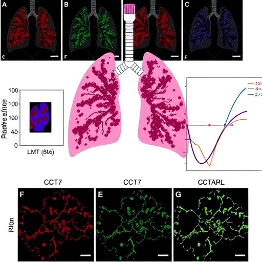
A protein better known for its day job inside the cell’s protein-folding assembly line may hold the key to predicting which patients with lung adenocarcinoma will fare worst, according to a new study published in Cancer Reports. Researchers report that CCT7, one of eight subunits of the chaperonin containing tailless complex polypeptide 1 (CCT)—also known as the TRiC ring complex—is consistently overproduced in lung adenocarcinoma (LUAD), the most common form of lung cancer, and that patients whose tumors carry high levels of the corresponding gene survive significantly shorter periods than those whose tumors do not. The findings, drawn from large public genomic datasets, an independent validation cohort and fresh tumor samples collected from patients undergoing lung surgery in China, position CCT7 as both a candidate diagnostic marker and a possible therapeutic target. In an era when lung cancer still kills more people than any other malignancy, the identification of a single molecular indicator that flags aggressive disease could sharpen how clinicians stage, monitor and treat the world’s deadliest cancer.
The scale of the problem provides the backdrop. Lung cancer is the most prevalent neoplasm worldwide and the leading cause of cancer-related death among women across North America, Europe and Australia. Pathologists divide the disease broadly into small cell lung cancer and non-small cell lung cancer (NSCLC), the latter accounting for roughly 85 percent of all cases and comprising two major subtypes: lung squamous cell carcinoma and lung adenocarcinoma. LUAD, which arises from mucus-producing glandular cells in the lung’s periphery, is the subtype most tightly linked to both smokers and never-smokers alike. Because early symptoms are notoriously non-specific—a persistent cough, mild breathlessness, unexplained fatigue—most patients are diagnosed only after the disease has advanced. The consequences are stark: individuals detected at stage IA enjoy a five-year survival rate approaching 90 percent, whereas those diagnosed at stage IV face a survival probability below 10 percent. Reliable early screening and accurate prognostic markers therefore rank among oncology’s most urgent unmet needs.
CCT7 belongs to one of biology’s most remarkable molecular machines. The CCT/TRiC complex is a large, barrel-shaped chaperonin located in the cytoplasm of eukaryotic cells, built from eight distinct protein subunits arranged in two stacked rings. Its job is to fold other proteins—most famously actin and tubulin, the structural building blocks of the cytoskeleton—into their functional three-dimensional shapes. Because tumor cells depend on rapid cytoskeletal remodeling to divide, invade surrounding tissue and migrate through the body, the CCT complex has long been suspected of fueling cancer progression. Earlier work showed that CCT subunits are overexpressed in head and neck squamous carcinoma, where higher levels correlate with worse patient outcomes, and that CCT7 in particular participates in autophagy and in the movement of fibroblasts, the connective-tissue cells that tumors recruit to build their supportive scaffolding. What remained unknown was whether CCT7 plays a comparable role in lung cancer—a question the Chinese team set out to answer with a combination of computational genomics and laboratory experiments.
The investigation began in silico. The researchers mined The Cancer Genome Atlas (TCGA), extracting gene-expression data and clinical records from 541 LUAD tumors and 59 matched non-cancerous lung samples, of which 508 cases carried sufficient survival information for outcome analysis. Patients were split into high- and low-CCT7 groups at the median expression value. To guard against dataset-specific artifacts, the team validated the findings in the Gene Expression Omnibus repository using the GSE31210 cohort—226 LUAD tumors and 20 matched normal tissues from patients with histologically confirmed primary disease. Protein-level confirmation came from the Human Protein Atlas immunohistochemistry archive, and then from the clinic: the researchers collected fresh tumor tissue and adjacent normal lung from 20 LUAD patients undergoing surgical resection at the First Affiliated Hospital of Guangxi Medical University between September and November 2023, none of whom had received prior chemotherapy, radiotherapy or other adjuvant treatment. Quantitative reverse-transcription polymerase chain reaction, using the standard 2−ΔΔCt method against a GAPDH reference gene, quantified CCT7 messenger RNA in every paired sample.
Every line of evidence converged on the same conclusion. In the TCGA cohort, CCT7 messenger RNA was markedly elevated in tumors relative to normal lung tissue (p < 0.001); the independent GSE31210 dataset reproduced the difference (p < 0.01); and the 20 fresh surgical sample pairs showed significant overexpression (p < 0.05). Immunohistochemical images from the Human Protein Atlas revealed visibly stronger CCT7 protein staining in tumor sections than in healthy tissue. Diagnostic potential was quantified with receiver operating characteristic (ROC) analysis, which measures how cleanly a marker separates diseased from healthy samples. CCT7 achieved an area under the curve (AUC) of 0.902 in TCGA and 0.722 in GSE31210—values indicating strong discriminatory power, particularly in the discovery cohort. An AUC of 1.0 represents perfect classification while 0.5 reflects chance performance alone; by that benchmark, the expression of a single gene distinguishes cancerous from non-cancerous lung tissue with an accuracy few accessible markers currently match.
CCT7’s signal extended beyond diagnosis into prognosis. Patients with stage III or IV disease expressed significantly more CCT7 than those with stage I or II tumors (p < 0.001); levels were also higher in larger primary tumors classified as T2, T3 or T4 compared with T1 lesions (p < 0.001), and in patients whose cancer had spread to regional lymph nodes (N1–N3) compared with node-negative cases (p < 0.001). Kaplan–Meier survival analysis, which tracks the proportion of patients remaining alive over time and compares curves with the log-rank test, showed that the high-CCT7 group in TCGA experienced significantly shorter overall survival (p = 0.031), a result independently confirmed in GSE31210 (p = 0.0022). To translate the finding into a practical bedside tool, the team constructed a nomogram—a graphical scoring chart—that integrates age, sex, TNM stage and CCT7 expression to predict one-, two-, three-, five- and ten-year survival. The model achieved a Harrell’s concordance index of 0.758, produced calibration curves closely matching observed outcomes, and returned AUC values of 0.646, 0.585 and 0.542 for the one-, three- and five-year predictions.
To understand what CCT7 is actually doing inside tumor cells, the researchers compared gene expression between high- and low-CCT7 tumors and identified 5,194 differentially expressed genes—4,095 upregulated and 1,099 downregulated—using stringent thresholds of at least a twofold change and an adjusted p-value below 0.05. Gene Ontology enrichment analysis pointed toward cytoskeleton organization, cell motility and microtubule-related processes, alongside molecular functions such as sodium and metal ion transmembrane transport. Kyoto Encyclopedia of Genes and Genomes pathway analysis linked the gene set to retinol metabolism, olfactory transduction and, notably, the metabolism of foreign compounds by cytochrome P450 enzymes—a detoxification system previously implicated in LUAD development. Gene Set Enrichment Analysis tied the program to broad biological regulation, cellular processes and multicellular organismal development. A protein–protein interaction network built with the STRING database revealed that CCT7 functionally connects with PHB, YWHAQ, RAN and CCT4, with PHB, RAN and CCNB1 emerging as hub genes. Each partner carries its own cancer pedigree: PHB is overexpressed in LUAD tissue, YWHAQ promotes invasion in non-small cell lung cancer, RAN activates the PI3K–AKT pathway to drive cell migration, and CCT4 participates in liver cancer and Wilms’ tumor biology.
Perhaps the most provocative findings concerned the tumor immune microenvironment—the constellation of immune cells that colonize tumors and shape their response to treatment. Using CIBERSORT, a computational algorithm that deconvolves bulk gene-expression data into the proportions of 22 immune cell types, the team found that high CCT7 expression correlated positively with activated memory CD4-positive T cells, CD8-positive T cells, T follicular helper cells and M1 macrophages, but negatively with regulatory T cells, monocytes, M2 macrophages, resting dendritic cells and resting mast cells. A parallel single-sample gene set enrichment analysis of 28 immune cell signatures confirmed broad associations across T cell, natural killer cell and dendritic cell lineages. Counterintuitively, ESTIMATE scoring—an algorithm that infers immune and stromal content from expression profiles—showed that tumors with high CCT7 carried lower immune and stromal scores and therefore greater tumor purity. Yet multiplex immunofluorescence staining of patient tissue demonstrated that CD8α, the hallmark marker of cytotoxic T lymphocytes, co-localized strongly with CCT7 in high-expressing tumors. The data point to a paradox: immune cells are present, but their functional state may be disrupted.
The mutational landscape added another layer. Patients in the high-CCT7 group carried a significantly higher tumor mutational burden—the total count of mutations within a tumor, a metric frequently used to predict responsiveness to immune checkpoint inhibitor therapy. Mutation waterfall plots generated with the maftools software package showed that mutations in TP53, CSMD3 and TTN each appeared in more than half of the high-CCT7 tumors. TP53, the so-called guardian of the genome, orchestrates DNA repair, apoptosis, cell-cycle arrest and cellular senescence; its inactivation is a hallmark of cancer and is associated with reduced survival and treatment resistance in lung adenocarcinoma. A high mutational burden ordinarily bodes well for immunotherapy, because accumulating mutations generate neoantigens the immune system can recognize. Yet the team also found that five immune checkpoint genes—VSIR, HAVCR2, SIRPA, BTLA and SIGLEC7—were expressed at lower levels in high-CCT7 tumors. These molecules normally restrain immune activity, and their depletion implies deeper disruption of immune homeostasis, potentially explaining why high-CCT7 patients present at more advanced stages with more lymph node metastases.
The authors conclude that CCT7 overexpression is a meaningful diagnostic and prognostic signal in LUAD and may represent a druggable vulnerability, given the chaperonin’s central role in cytoskeletal dynamics. The study’s design also defines its limits: bulk sequencing cannot resolve whether elevated CCT7 originates from tumor cells, stromal cells or infiltrating immune cells, and the precise mechanism by which a protein-folding factor modulates checkpoint expression remains to be defined. The nomogram, although internally well calibrated, will require prospective validation in independent cohorts before clinical adoption. Even so, the convergence of computational prediction, independent database validation, fresh patient samples and protein-level imaging gives the finding unusual weight. If future functional studies confirm that silencing CCT7 impairs LUAD growth or restores immune competence, this humble folding assistant of actin and tubulin could graduate from statistical biomarker to therapeutic target—offering a new lever against a disease that still claims more lives worldwide than any other cancer.
Cite Scienmag News
Nathaniel Bowman. (August 30, 2026). CCT7 Expression Emerges as a Prognostic Marker in Lung Adenocarcinoma. Scienmag. https://scienmag.com/cct7-expression-emerges-as-a-prognostic-marker-in-lung-adenocarcinoma/
Nathaniel Bowman. "CCT7 Expression Emerges as a Prognostic Marker in Lung Adenocarcinoma." Scienmag, 30 August 2026, https://scienmag.com/cct7-expression-emerges-as-a-prognostic-marker-in-lung-adenocarcinoma/. Accessed 3 September 2026.
Nathaniel Bowman. "CCT7 Expression Emerges as a Prognostic Marker in Lung Adenocarcinoma." Scienmag. August 30, 2026. https://scienmag.com/cct7-expression-emerges-as-a-prognostic-marker-in-lung-adenocarcinoma/


