


Chiba, Japan—Hallucinations and delusions that first emerge in midlife or old age may sometimes reflect hidden neurodegenerative changes rather than a purely psychiatric disorder, according to a new brain-imaging study published in Molecular Psychiatry. Researchers using positron emission tomography (PET) found that abnormal tau protein accumulation was far more common in people with late-onset psychosis than in healthy older adults. The findings offer some of the clearest evidence yet that psychotic symptoms appearing after age 40 can be associated with biological changes linked to dementia.
The study examined 37 patients whose psychosis began after the age of 40 and compared them with 47 healthy older adults. The participants underwent two types of PET scanning: one designed to detect amyloid plaques, a hallmark of Alzheimer’s disease, and another using QST’s florzolotau (18F) tracer to visualize tau pathology in the living brain. Tau is a structural protein that normally helps stabilize neurons, but in several neurodegenerative diseases it becomes abnormally modified, accumulates inside nerve cells and disrupts their function.
The difference between the two groups was striking. Tau PET scans were positive in approximately 65% of patients with late-onset psychosis, compared with only 15% of healthy controls. Amyloid PET positivity was also substantially higher among patients, occurring in 35% compared with 2% of controls. These results suggest that a significant proportion of people who develop hallucinations or delusions later in life may carry measurable molecular signatures of neurodegeneration, even when dementia has not yet been clearly diagnosed.
The researchers emphasize that late-onset psychosis is not a single biological condition. Patients can experience similar symptoms while having very different disease processes in the brain. Some participants showed both amyloid and tau accumulation, a pattern consistent with Alzheimer’s disease. Others had tau buildup without detectable amyloid plaques, pointing toward non-Alzheimer’s forms of tau-related neurodegeneration. This distinction is important because amyloid-negative tau pathology can occur in several other neurological disorders and may follow different clinical courses.
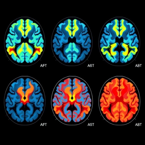
The brain scans also revealed that tau was not distributed uniformly across patients. In several amyloid-negative cases, accumulation was particularly evident in posterior regions, including the parietal and occipital lobes. These areas contribute to attention, visual interpretation, spatial processing, judgment and the integration of information from different parts of the brain. Disruption in these systems could help explain why some patients develop false perceptions, unusual beliefs or difficulty interpreting their surroundings.
One representative set of images showed four distinct patterns of tau deposition. The first case displayed an amyloid-positive pattern associated with Alzheimer’s disease, while the remaining cases were amyloid-negative and appeared to represent other types of tau pathology. White arrowheads in the images marked areas of abnormal tau accumulation. Together, the scans illustrate why late-onset psychosis can be difficult to classify using symptoms alone: identical hallucinations or delusions may arise from different molecular changes and different networks of affected brain regions.
The study also identified a relationship between tau burden and cognitive performance. Among patients who were amyloid-positive, greater tau accumulation in the parietal lobe was associated with poorer executive function. Executive abilities include planning, shifting attention, monitoring behavior and solving problems. Although the study does not prove that tau directly causes psychosis, the association suggests that regional neurodegeneration may influence both cognitive decline and the emergence of psychiatric symptoms.
For clinicians, the findings could eventually change how new-onset psychosis in older adults is investigated. Psychiatric assessment remains essential, but symptoms alone may not reveal whether a patient has an underlying neurodegenerative disorder. PET imaging and other biological tests could help identify Alzheimer’s-related disease, non-Alzheimer’s tau pathology or a condition without substantial protein accumulation. Earlier identification may allow patients and families to receive more appropriate counseling, monitoring and treatment, while also helping researchers design clinical trials for biologically defined groups.
The researchers caution that the results should not be interpreted as evidence that every older person with hallucinations or delusions has dementia. The sample was relatively small, and PET positivity does not automatically predict when or whether an individual will develop significant cognitive impairment. Further studies involving larger and more diverse populations will be needed to determine how tau patterns evolve over time and whether they can predict treatment response. Nevertheless, the work moves late-onset psychosis closer to the era of molecular diagnosis, suggesting that what appears to be a psychiatric mystery may, in many cases, be a visible process unfolding inside the aging brain.
News Publication Date: 2 August 2026
Web References: https://doi.org/10.1038/s41380-026-03749-3
References: Molecular Psychiatry, DOI: 10.1038/s41380-026-03749-3
Cite Scienmag News
Cassandra Pierce. (August 4, 2026). Brain scans detect tau accumulation in late-onset psychosis. Scienmag. https://scienmag.com/brain-scans-detect-tau-accumulation-in-late-onset-psychosis/
Cassandra Pierce. "Brain scans detect tau accumulation in late-onset psychosis." Scienmag, 4 August 2026, https://scienmag.com/brain-scans-detect-tau-accumulation-in-late-onset-psychosis/. Accessed 4 September 2026.
Cassandra Pierce. "Brain scans detect tau accumulation in late-onset psychosis." Scienmag. August 4, 2026. https://scienmag.com/brain-scans-detect-tau-accumulation-in-late-onset-psychosis/


