Long before an individual forgets a name or loses their keys, the brain may already be quietly changing. One of the earliest and most elusive signals of impending Alzheimer’s disease is subjective cognitive decline, a stage in which people report that their memory or thinking feels worse than before, even though standard neuropsychological tests still come back normal. Because the underlying brain alterations at this stage are subtle, diffuse, and highly variable from person to person, clinicians have struggled to detect SCD reliably with conventional imaging analysis. A new study published in BMC Medical Imaging suggests that a carefully engineered hybrid of deep learning and radiomics can pull this faint signal out of ordinary T1-weighted MRI scans, achieving an accuracy of nearly 89 percent in distinguishing people with SCD from healthy controls.
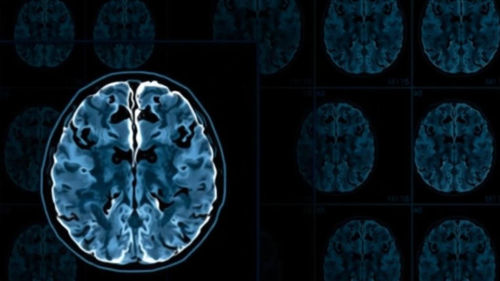
The research, led by Zhuoyuan Li, Xingxing Cen, Junyi Yuan, and Linhui Mi of the Information Center at Shanghai Chest Hospital, Shanghai Jiao Tong University School of Medicine, drew on data from the Alzheimer’s Disease Neuroimaging Initiative, one of the most extensively characterized longitudinal studies of brain aging in the world. The team assembled 234 normal controls and 229 individuals with subjective cognitive decline, all of whom had undergone structural T1-weighted magnetic resonance imaging. The MRI data were preprocessed using standardized procedures to ensure that differences in scanner settings and image quality would not contaminate the analysis. From this single, widely available imaging modality, the researchers extracted two fundamentally different kinds of information: hand-crafted radiomics features and deep features learned automatically by a vision transformer.
Radiomics is the practice of converting medical images into large panels of quantitative descriptors. Rather than asking a radiologist to eyeball the scan, radiomics computes hundreds of numerical measurements that capture texture, shape, and intensity patterns invisible to the human eye. In this study, the feature families included gray level co-occurrence matrices, gray level run length matrices, gray level size zone matrices, and gray level dependence matrices, each of which quantifies different aspects of how pixel intensities are distributed and correlated across the brain. Filters such as the Laplacian of Gaussian were applied to highlight structures at particular spatial scales. These features are interpretable and computationally cheap, but they are limited to patterns that a human engineer thought to measure in advance.
Deep learning takes the opposite approach. A vision transformer, or ViT, is a neural network architecture that divides an image into patches and uses self-attention mechanisms to model relationships between distant parts of the image. Unlike convolutional networks, which build up understanding through local filters layer by layer, transformers can in principle connect information from any region of the brain to any other, making them well suited to detecting the widespread, low-amplitude changes that characterize early neurodegeneration. The deep features extracted by the ViT encode patterns that no one explicitly designed, but they are opaque and may overlap with or even duplicate information already captured by the radiomics panel.
The central innovation of the study is the Hybrid Vision-Radiomics Aggregation framework, or HVRA, which the researchers designed to merge these two complementary views of the brain without letting redundancy drown out the signal. The first step is feature selection based on the minimum redundancy maximum relevance criterion, a well-established method that scores candidate features by how strongly they relate to the classification target while penalizing features that carry information already provided by others. By running mRMR selection repeatedly, the team identified a set of consensus features that appeared consistently across iterations, indicating that they were stable rather than statistical flukes.
After selection, the surviving radiomics features and the deep learning features were combined through weighted feature concatenation, in which each feature stream receives a learned weight reflecting its contribution to the classification task. This design distinguishes HVRA from cruder fusion strategies. Direct concatenation simply stacks both feature vectors together, allowing redundant or noisy features to dilute the informative ones. Learnable fusion approaches, in contrast, hand the entire integration problem to a neural network, which can overfit when training data are limited. HVRA sits between these extremes: it prunes the radiomics side with a principled statistical criterion and then lets a modest weighting mechanism balance the two modalities. The result, according to the authors, is a model that extracts the best of both worlds while remaining robust to the moderate sample sizes typical of clinical neuroimaging studies.
The performance numbers are striking for a task this difficult. Evaluated with a repeated cross-validation strategy inside the ADNI cohort, the HVRA model achieved an accuracy of 88.98 percent with a standard deviation of 3.42 percent, and an area under the receiver operating characteristic curve of 0.941. The AUC, which measures how well a model ranks positive cases above negative ones across all possible decision thresholds, indicates that the framework separates SCD from normal controls with high confidence. Crucially, HVRA outperformed every unimodal baseline, meaning that neither deep features nor radiomics features alone could match the integrated model. It also beat conventional fusion strategies, including direct concatenation and learnable fusion, confirming that the structured aggregation step is what drives the improvement rather than the raw availability of two feature types.
Perhaps the most scientifically intriguing result concerns the four consensus features that the selection procedure repeatedly converged upon. These features were not only stable across cross-validation runs; they also showed significant correlations with participants’ cognitive performance, as measured by neuropsychological instruments. In other words, the quantitative imaging signatures that the algorithm found most useful for detecting SCD also tracked how well individuals actually performed on cognitive testing. This alignment between machine-derived features and clinical measures suggests that the model is not latching onto scanner artifacts or demographic confounds, but is instead capturing biologically meaningful brain alterations associated with the earliest symptomatic complaints of Alzheimer’s disease. The authors note that these features may hold potential relevance for characterizing SCD-related brain changes, though they caution that the findings come from internal validation within a single cohort.
The clinical implications are considerable. Subjective cognitive decline is regarded as a key preclinical stage of Alzheimer’s disease, and identifying it accurately opens a window for intervention years or even decades before dementia manifests. Current approaches to early detection often rely on positron emission tomography tracers for amyloid-β or on invasive and expensive cerebrospinal fluid assays, neither of which is practical for broad population screening. A method that works from a standard T1-weighted MRI, a scan already performed in countless clinics, could dramatically lower the barrier to early identification. The authors emphasize that deep learning has become a promising strategy for improving disease feature extraction in medical imaging, but that its application to neurodegenerative diseases has remained limited precisely because the relevant changes are so subtle; a hybrid framework that amplifies weak signals may help overcome that barrier.
As with any machine learning study in medicine, important caveats remain. The model was validated internally within the ADNI cohort using repeated cross-validation, a rigorous approach for estimating performance, but external validation on independent datasets from different scanners, populations, and geographic regions will be essential before the method can move toward clinical use. The ADNI cohort itself is carefully curated, and real-world clinical populations are typically noisier and more heterogeneous. Nevertheless, the study demonstrates a clear principle: when deep learning and radiomics are integrated thoughtfully, with explicit attention to redundancy and relevance, the combination can detect brain states that neither approach reveals alone. For the millions of people who sense that something has changed in their own minds long before any test can confirm it, that principle may one day translate into earlier answers, earlier monitoring, and a genuine head start against Alzheimer’s disease.
Subject of Research: Multimodal deep learning and radiomics for early detection of subjective cognitive decline on MRI
Article Title: Multimodal integration of deep learning and radiomics enables early detection of subjective cognitive decline
Article References: Li, Z., Cen, X., Yuan, J., & Mi, L. (2026). Multimodal integration of deep learning and radiomics enables early detection of subjective cognitive decline. BMC Medical Imaging. https://doi.org/10.1186/s12880-026-02893-0
Image Credits: AI Generated
DOI: 10.1186/s12880-026-02893-0
Keywords: subjective cognitive decline, Alzheimer's disease, radiomics, deep learning, vision transformer, MRI, multimodal fusion, feature selection, machine learning, ADNI, early detection, neurodegeneration
Cite Scienmag News
Cassandra Pierce. (October 11, 2026). AI Spots the Earliest Whisper of Alzheimer’s Disease in Routine Brain Scans. Scienmag. https://scienmag.com/ai-spots-the-earliest-whisper-of-alzheimers-disease-in-routine-brain-scans/
Cassandra Pierce. "AI Spots the Earliest Whisper of Alzheimer’s Disease in Routine Brain Scans." Scienmag, 11 October 2026, https://scienmag.com/ai-spots-the-earliest-whisper-of-alzheimers-disease-in-routine-brain-scans/. Accessed 11 October 2026.
Cassandra Pierce. "AI Spots the Earliest Whisper of Alzheimer’s Disease in Routine Brain Scans." Scienmag. October 11, 2026. https://scienmag.com/ai-spots-the-earliest-whisper-of-alzheimers-disease-in-routine-brain-scans/