


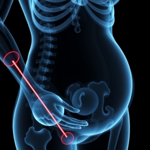
Unfractionated heparin, one of the oldest weapons in medicine’s arsenal against blood clots, may be quietly draining strength from the skeletons of the very pregnant women it is meant to protect. That is the provocative conclusion of a new study from the University of Tokyo, published in the journal Reproductive Sciences, in which an artificial intelligence system read ordinary preoperative chest X-rays and found that expectant mothers exposed to the drug had significantly lower estimated bone mineral density at both the lumbar spine and the femoral neck than women who underwent cesarean section without it. The finding matters because the gold-standard tool for measuring bone density, dual-energy X-ray absorptiometry, is avoided during pregnancy over concerns about fetal radiation exposure, leaving one of the most feared complications of anticoagulant therapy nearly invisible at precisely the moment it matters most. The Japanese team’s workaround — extracting bone data with AI from images already taken for surgical safety — may finally open a window onto this hidden territory.
Heparin occupies an awkward position in modern obstetrics. Pregnancy itself tilts the blood’s clotting balance toward danger: venous stasis, vessel injury at delivery and a surge in clotting factors make venous thromboembolism a leading cause of maternal death and long-term disability in high-income countries. For some women, anticoagulation is not optional — those with antiphospholipid syndrome, in whom heparin combined with low-dose aspirin has repeatedly been shown to reduce recurrent miscarriage; patients with inherited thrombophilias; women with mechanical heart valves; and those who suffer an acute clot during the pregnancy itself. Low-molecular-weight heparin has become the modern mainstay because it can be injected once or twice daily, does not measurably cross the placenta and carries a comparatively modest skeletal risk. Unfractionated heparin, however, has not vanished from the ward. It acts instantly, its effect is fully reversible with the antidote protamine, and it remains the agent of choice when kidney function is severely impaired or when urgent procedures or bleeding risks demand rapid control. In precisely these situations, a substantial number of pregnancies remain exposed to the older drug’s notorious skeletal side effects.
The link between heparin and bone loss was recognized decades ago, when patients receiving prolonged therapy for thrombotic disease began presenting with vertebral and hip fractures, and heparin-induced osteoporosis entered the textbooks as a feared complication of long-term treatment. Quantifying that loss during pregnancy, however, has always collided with a hard technical barrier. Dual-energy X-ray absorptiometry, which separates bone from soft tissue using two distinct X-ray energies and reports areal density in grams per square centimeter, is almost never performed on pregnant patients; even though the fetal radiation dose is minuscule, elective exposure is routinely deferred. Radiation-free surrogates such as quantitative ultrasound offer only limited diagnostic agreement with DXA at the spine, hip and forearm, according to validation studies cited by the researchers. The result is a scientific blind spot around pregnancy- and lactation-associated osteoporosis, a rare but devastating condition in which previously healthy young women sustain fragility fractures of the vertebrae in late pregnancy or during breastfeeding — injuries often dismissed as ordinary back pain until imaging reveals collapsed vertebral bodies.
The Tokyo collaboration, which unites the University of Tokyo’s departments of obstetrics and gynecology, orthopedic surgery and preventive medicine with engineers at the technology company KYOCERA, engineered its way around the barrier with deep learning. The group had previously developed and validated an artificial intelligence-assisted diagnostic system, described in the Journal of Orthopaedic Research, that estimates bone mineral density at the lumbar spine and femoral neck from plain radiographs. Trained on large collections of X-ray images paired with conventional DXA measurements, the neural network learns to detect the fine-scale texture and architectural signatures of trabecular bone — radiographic patterns far too subtle for the human eye that nevertheless correlate tightly with true density — and converts them into an estimated bone mineral density, or eBMD. The same platform has since been used to evaluate bone changes in postmenopausal women after total hip replacement surgery. Its seductive promise is opportunistic screening: any radiograph acquired for an unrelated clinical reason becomes, with zero additional radiation, cost or scheduling, a quantitative test of skeletal health.
In the new work, the team pointed that promise at the delivery room. Between April 2013 and October 2023, the researchers assembled a retrospective cohort from their institution comprising 86 pregnant women who had received unfractionated heparin therapy during pregnancy and 213 women who underwent cesarean delivery without any medication, all of whom had usable preoperative chest radiographs obtained before surgery. The AI system assigned each participant estimated bone mineral density values for the lumbar spine and the femoral neck. The two groups were then compared with univariate statistics, followed by multivariate regression models adjusted for relevant covariates such as body mass index, in order to isolate the contribution of heparin itself from other influences on the maternal skeleton. The study received ethics approval from the University of Tokyo’s institutional review board and was supported by Japan’s Ministry of Health, Labour and Welfare, the Japan Society for the Promotion of Science and KYOCERA Corporation.
The results were strikingly consistent. Compared with the 213 controls, the 86 heparin-exposed women showed significantly lower estimated bone mineral density at both skeletal sites examined. In the multivariate analysis, unfractionated heparin exposure emerged as an independent factor associated with reduced bone density at the lumbar spine, meaning the association survived statistical adjustment for the other variables measured. Body mass index correlated with eBMD in both groups, but the interaction proved clinically poignant: underweight women, classified according to body mass index thresholds established for Asian populations, displayed significantly lower estimated density, and the deficit was most pronounced among underweight women who had also received heparin. The double burden of scant body reserves and anticoagulant exposure appears to fall hardest on the maternal skeleton — a sobering pattern, given that many of the women placed on heparin for recurrent pregnancy loss are themselves young and slim. Notably, the researchers found no significant correlation between the total cumulative heparin dose and eBMD, a puzzle with important mechanistic implications.
Equally telling was where the loss concentrated. The lumbar spine proved more vulnerable than the femoral neck, a site-specificity the authors highlight in their conclusions. Skeletal biology offers a ready explanation. Vertebral bodies are packed with trabecular bone, the honeycombed interior scaffolding whose vast surface area and rapid remodeling turnover make it the body’s first responder to metabolic stress; the femoral neck, by contrast, is sheathed in a thicker mantle of slow-turning cortical bone. Insults to the skeleton — lactation, glucocorticoid therapy and now, apparently, heparin — therefore register earliest and most severely in the spine, which is also where fractures cluster in pregnancy-associated osteoporosis. Timing compounds the physiological squeeze. The fetal skeleton accumulates roughly thirty grams of calcium, the great majority of it during the final trimester, drawing on the mother’s markedly increased intestinal absorption and, when that supply falls short, on resorption of her own bone. Any drug that further erodes that reserve exploits a system already running near its limit.
How heparin wounds bone has been investigated for half a century without full resolution, but the mechanisms converge on a two-pronged assault: suppression of osteoblasts, the cells that build new bone, alongside promotion of osteoclast-driven resorption of existing matrix, with heparin molecules also capable of binding calcium and perturbing vitamin D metabolism. Low-molecular-weight heparins were developed partly to shed this toxicity, and the comparative clinical evidence has been mixed — a randomized substudy of long-term dalteparin in pregnancy found no decrease in bone mineral density, while observational cohorts of women on prolonged low-molecular-weight therapy have documented measurable loss, and studies in dialysis patients suggest both heparin classes can affect the skeleton. Against that backdrop, one detail of the new study stands out: the total cumulative heparin dose showed no significant correlation with estimated bone density. The absence of a dose-response relationship hints that exposure itself, or its timing early in gestation, may matter more than the sheer number of units administered — while also leaving room for confounding by indication, since women who require unfractionated heparin may differ fundamentally from those who do not.
The authors are appropriately measured about the limits of a retrospective, single-center design. The eBMD values are algorithmic estimates rather than direct DXA measurements; the chest radiographs were acquired for surgical preparation, not densitometry; no fracture outcomes or postpartum follow-up data were reported; and observational associations, however carefully adjusted, cannot by themselves establish causation. Yet the internal coherence of the findings is difficult to dismiss: the heparin effect survived multivariate adjustment, matched the predicted anatomical pattern of trabecular susceptibility, interacted with low body weight in a biologically plausible direction and aligns with a long experimental literature on heparin’s suppression of bone-forming cells. For a complication this rarely quantified, carefully adjusted observational evidence of this kind meaningfully shifts the burden of proof — and argues for prospective studies in which women on heparin are tracked with both AI-estimated and conventional measurements after delivery.
The implications stretch well beyond one drug. The study sketches a template for pregnancy-safe skeletal surveillance: mining radiographs that already exist in medical records, translating them through AI into quantitative density estimates and using the results to stratify risk before a fracture ever occurs. The researchers conclude that the estimation system may provide a safe, accessible and effective method for early detection and risk stratification of pregnancy-associated bone loss, supporting future clinical management of at-risk populations — beginning, perhaps, with underweight women on heparin, who might warrant closer attention to calcium and vitamin D intake, carefully weighed anticoagulant selection and postpartum densitometry once fetal radiation ceases to be a concern. If the approach is validated prospectively, it could generalize far beyond this single scenario, converting the millions of X-rays taken each year into an untapped archive of skeletal information. For now the message is narrower but urgent: a treatment given to safeguard two lives may quietly tax one of them, and algorithms that read the images are beginning to keep score.
Cite Scienmag News
Harold Sullivan. (August 29, 2026). AI Radiographic Analysis Links Heparin in Pregnancy to Reduced Maternal Bone Density. Scienmag. https://scienmag.com/ai-radiographic-analysis-links-heparin-in-pregnancy-to-reduced-maternal-bone-density/
Harold Sullivan. "AI Radiographic Analysis Links Heparin in Pregnancy to Reduced Maternal Bone Density." Scienmag, 29 August 2026, https://scienmag.com/ai-radiographic-analysis-links-heparin-in-pregnancy-to-reduced-maternal-bone-density/. Accessed 3 September 2026.
Harold Sullivan. "AI Radiographic Analysis Links Heparin in Pregnancy to Reduced Maternal Bone Density." Scienmag. August 29, 2026. https://scienmag.com/ai-radiographic-analysis-links-heparin-in-pregnancy-to-reduced-maternal-bone-density/


