


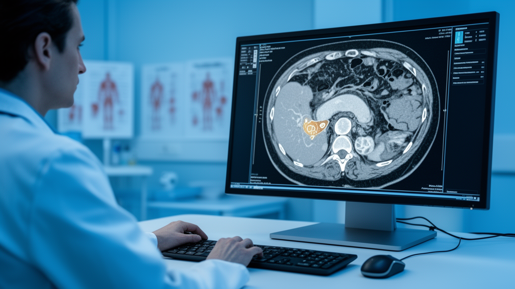
For patients diagnosed with a pancreatic neuroendocrine neoplasm, one of the most consequential questions is also one of the hardest to answer before surgery: is the tumor a relatively indolent neuroendocrine tumor, or the far more aggressive neuroendocrine carcinoma? The distinction between these two entities, collectively known as pancreatic neuroendocrine neoplasms, shapes everything from the extent of resection to the use of adjuvant therapy. Yet on a conventional CT scan, the two can look deceptively similar. A new study published in BMC Medical Imaging suggests that a fusion of advanced spectral CT physics, radiomics, and classical statistical modeling may finally give radiologists a reliable preoperative answer.
The research, led by Jiajia Shi and Yunjin Chen of the First Affiliated Hospital of Zhengzhou University together with colleagues across multiple Chinese institutions, enrolled 128 patients who had undergone contrast-enhanced dual-layer spectral detector CT and whose diagnoses were confirmed by pathology. Of these, 86 carried pancreatic neuroendocrine tumors while 42 had the more malignant carcinomas. The cohort was randomly split into a training set of 89 patients and a validation set of 39, in a seven-to-three ratio, allowing the investigators to build their models on one group and test them honestly on another.
The technological heart of the study is dual-layer spectral detector CT, a scanner architecture that separates the energies of X-ray photons at the detector itself rather than requiring rapid voltage switching. This capability allows the reconstruction of virtual monoenergetic images, which simulate how tissue would appear if imaged with a single X-ray energy. The team focused on images reconstructed at 40 kiloelectron volts during the venous phase of contrast enhancement. At this low energy, iodine contrast attenuation is dramatically amplified, exaggerating subtle differences in how the two tumor types take up contrast agent and exposing texture information that conventional polychromatic images wash out.
From these 40 keV images, the researchers extracted radiomics features, a family of quantitative descriptors that capture first-order statistics of pixel intensity, spatial relationships between neighboring voxels, and higher-order texture patterns invisible to the human eye. The underlying premise is that tumor biology leaves fingerprints in image texture: differences in cell density, fibrosis, necrosis, and vascularity between well-differentiated tumors and poorly differentiated carcinomas translate into measurable mathematical signatures. Combined with conventional CT morphological features and spectral parameters such as iodine concentration and effective atomic number, these features formed the raw material for a series of logistic regression models.
The investigators constructed four competing models in a deliberate escalation of complexity. The first relied solely on conventional CT features, the second added dual-layer spectral detector CT parameters, the third used only the radiomics features derived from the 40 keV virtual monoenergetic images, and the fourth integrated all three data streams into a single combined model. Performance was quantified using the area under the receiver operating characteristic curve, a standard metric expressing how well a model separates the two diagnostic classes across all possible decision thresholds.
The results were striking. In the training cohort, the combined model achieved an area under the curve of 0.956, substantially outperforming the conventional CT model at 0.794, the spectral CT model at 0.835, and the radiomics model at 0.871. DeLong’s test, a statistical procedure for comparing correlated areas under the curve, confirmed that these differences were significant, with all comparisons yielding p values below 0.05. In the validation cohort, the combined model retained strong performance at 0.905, though the gaps to the simpler models, which scored 0.787, 0.805, and 0.811 respectively, were no longer statistically significant, a common pattern when sample sizes are modest.
Beyond raw discrimination, the team interrogated the combined model with decision curve analysis and calibration assessment. Decision curve analysis evaluates the net clinical benefit of acting on a model’s predictions across a range of threshold probabilities, asking whether the model would lead to better decisions than treating all patients or none. Calibration analysis examines whether predicted probabilities match observed frequencies. Both analyses supported the robustness and favorable goodness-of-fit of the combined model, indicating that its high area under the curve was not a statistical artifact but reflected genuinely useful predictive structure.
To translate the model into clinical practice, the researchers plotted a nomogram, a graphical scoring tool that assigns weighted points to each predictive variable and sums them into a probability estimate. Nomograms occupy a beloved niche in oncology because they make complex multivariable models legible at the bedside: a clinician can read off a patient’s feature values, tally the points, and obtain an individualized risk estimate without any computation. In this case, the nomogram distills conventional CT findings, spectral parameters, and radiomics signatures into a single preoperative probability that a pancreatic neuroendocrine neoplasm is a tumor rather than a carcinoma.
The clinical stakes of such a tool are considerable. Pancreatic neuroendocrine tumors and neuroendocrine carcinomas demand different surgical strategies and different postoperative pathways, and preoperative uncertainty often forces clinicians to plan for the worst case. A validated noninvasive discriminator could refine surgical planning, inform the aggressiveness of lymph node dissection, and help select patients for neoadjuvant approaches. Because the entire workflow relies on images already acquired during standard contrast-enhanced CT examinations, the marginal cost and radiation burden to the patient are essentially zero, a significant advantage over additional imaging or biopsy.
The authors are appropriately measured about the limits of their work. The study was retrospective, conducted in accordance with the Declaration of Helsinki with ethics approval from the First Affiliated Hospital of Zhengzhou University, and drew on a relatively small single-region cohort of 128 patients. The validation set of 39 patients, while sufficient to demonstrate promise, cannot establish superiority over simpler models with statistical confidence, as the non-significant DeLong comparisons in that cohort make clear. The team concludes that the combined model shows promising performance that may aid clinical decision-making, but that its superiority requires further validation in external independent cohorts. If larger multi-center studies replicate these findings, spectral CT radiomics nomograms could become a standard preoperative instrument, turning routine CT scans into molecularly informative diagnostics for one of the pancreas’s most diagnostically treacherous tumor families.
Subject of Research: Preoperative differentiation of pancreatic neuroendocrine tumors from carcinomas using spectral CT radiomics and a nomogram
Article Title: A spectral CT radiomics nomogram for preoperative discrimination between pancreatic neuroendocrine tumors and carcinomas
Article References: A spectral CT radiomics nomogram for preoperative discrimination between pancreatic neuroendocrine tumors and carcinomas. (n.d.). https://doi.org/10.1186/s12880-026-02869-0
Image Credits: AI Generated
DOI: 10.1186/s12880-026-02869-0
Keywords: pancreatic neuroendocrine neoplasms, spectral CT, dual-layer detector CT, radiomics, nomogram, virtual monoenergetic images, machine learning, medical imaging, pancreatic cancer, logistic regression, diagnostic imaging, BMC Medical Imaging
Cite Scienmag News
Ophelia Keating. (October 14, 2026). AI-Powered CT Scan Model Distinguishes Pancreatic Tumors Before Surgery. Scienmag. https://scienmag.com/ai-powered-ct-scan-model-distinguishes-pancreatic-tumors-before-surgery/
Ophelia Keating. "AI-Powered CT Scan Model Distinguishes Pancreatic Tumors Before Surgery." Scienmag, 14 October 2026, https://scienmag.com/ai-powered-ct-scan-model-distinguishes-pancreatic-tumors-before-surgery/. Accessed 14 October 2026.
Ophelia Keating. "AI-Powered CT Scan Model Distinguishes Pancreatic Tumors Before Surgery." Scienmag. October 14, 2026. https://scienmag.com/ai-powered-ct-scan-model-distinguishes-pancreatic-tumors-before-surgery/


