


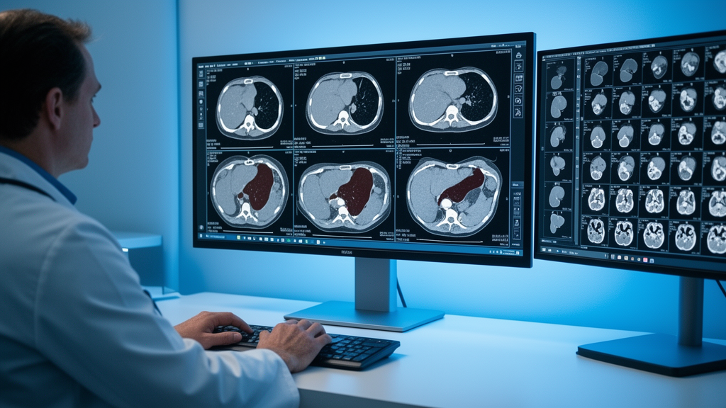
Deep-learning models that outline liver tumors on computed tomography scans promise to speed up diagnosis and treatment planning, but they share a stubborn weakness: they expect the world to look like their training data. In clinical practice, that expectation is routinely broken. A patient may arrive with only a non-contrast scan, or with contrast-enhanced images from some phases but not others, and the annotations used to train the model may cover only a subset of the available phases. A new study published in BMC Medical Imaging tackles exactly this gap, reporting a segmentation network called MFANet that was designed from the ground up to tolerate missing CT phases and heterogeneous, partially labeled datasets from multiple sources.
The work, led by Yue Zhang, JunXuan Wu, DeJian Xing and Shumei Zhang of the School of Computer Science and Artificial Intelligence at Northeast Forestry University in Harbin, China, was published on 14 October 2026 as an open-access research article. The team describes MFANet as a missing-phase-tolerant multi-source frequency–anatomy network, a name that hints at its two central ideas. The first is a masked phase fusion strategy, in which the model learns to combine information from whichever CT phases are available for a given patient while gracefully handling the absence of others. The second is a pair of separate decoders, one dedicated to the liver and one to tumors, allowing the network to optimize each task with its own anatomical and textural priorities rather than forcing a single output head to serve two very different structures.
The evaluation was deliberately rigorous. The researchers assembled five public datasets and split them at the patient level into a development pool of 1,029 cases and 260 within-source test cases, ensuring that no patient appeared in both training and testing. They then added an independent external dataset: a four-phase contrast-enhanced CT collection from Chongqing Yubei District People’s Hospital comprising 361 patients, of whom 278 had primary liver cancer. The remaining 83 patients were cancer-free controls, and the authors excluded them from tumor evaluation. Five competing segmentation models were given exactly the same phase information and label supervision as MFANet, so that any performance difference could be attributed to architecture rather than to unequal inputs.
The primary endpoint was the equal-source mean patient-level tumor Dice score across the five within-source test cohorts. The Dice score, a standard metric in medical image segmentation, measures the overlap between the model’s predicted tumor region and the expert-drawn ground truth, ranging from 0 for no overlap to 1 for perfect agreement. MFANet achieved a mean tumor Dice of 80.84 percent, compared with 79.84 percent for STU-Net-B, the strongest of the five comparators. The paired difference of 1.00 percentage point came with a 95 percent confidence interval of 0.38 to 1.59, which the researchers assessed using a source-stratified bootstrap. That statistical approach resamples the data within each source dataset, protecting the result from being driven by any single cohort. The interval lying entirely above zero indicates a consistent, if modest, advantage for MFANet.
Small lesions are where liver tumor segmentation is hardest and where the clinical stakes are arguably highest, because tiny nodules are easy for both radiologists and algorithms to overlook. In an exploratory analysis that required a lesion-level Dice of at least 0.10 to count a lesion as detected, MFANet reached a sensitivity of 61.2 percent for tumors smaller than 10 millimeters, against 56.5 percent for STU-Net-B. That gap of nearly five percentage points on sub-centimeter lesions suggests the architecture’s frequency-and-anatomy design may be particularly helpful for the fine-grained texture and shape cues that distinguish small tumors from surrounding liver tissue, although the authors characterize this analysis as exploratory rather than a definitive claim.
To probe how much each imaging phase actually matters, the team ran an ablation in which the portal-venous phase was removed from the input. On the same 168-case subset, Dice scores dropped for all six models tested. Notably, the researchers did not establish a between-model difference in the size of that decline, meaning they could not show that MFANet degrades more gracefully than its competitors when this diagnostically rich phase is absent. The result underscores a practical reality: contrast-enhanced phases, especially the portal-venous phase in which liver tumors typically stand out most clearly, carry substantial information that no current architecture can fully reconstruct from a non-contrast scan alone.
External validation provided a second, harder test. On the independent primary liver cancer contrast-enhanced CT dataset, MFANet achieved a tumor Dice of 77.10 percent among the 278 cancer cases, compared with 75.92 percent for STU-Net-B. Performance on data from an entirely different hospital, scanner fleet and imaging protocol is usually lower than on internal test sets, and the roughly four-point drop from the within-source average here is consistent with that pattern. Still, the fact that MFANet retained its edge over the strongest comparator on out-of-distribution data strengthens the case that the improvement is not an artifact of overfitting to the public benchmarks.
The authors are careful about the limits of their findings, and their candor is worth emphasizing. They conclude that MFANet modestly improved tumor segmentation under matched inputs and supervision in the tested cohorts, but they state plainly that its clinical value and its performance on naturally incomplete external scans and on cancer-free controls remain unknown. The external dataset, while independent, was curated for the study; real-world hospital archives contain scans with unpredictable combinations of phases, variable reconstruction kernels and artifacts that no public benchmark fully reproduces. The exclusion of the 83 control patients from tumor evaluation also means the study says nothing about how such a model behaves in patients without tumors, a scenario that matters for screening applications where false positives carry real costs.
The study’s methodology reflects a broader shift in how medical imaging AI is evaluated. Rather than reporting a single headline number on one benchmark, the team used patient-level partitions across five heterogeneous sources, matched inputs for all comparators, a prespecified primary endpoint, and bootstrap confidence intervals stratified by source. Secondary metrics such as the 95th-percentile Hausdorff distance and normalized surface Dice, which capture how far predicted boundaries stray from true ones, complement the overlap-based Dice score. The authors also report expected calibration error, a measure of how well the model’s confidence aligns with its actual accuracy, an increasingly important consideration for tools intended to support clinical decisions.
For clinicians and researchers, the message is twofold. Architectural choices that explicitly model phase availability and separate organ-level from lesion-level decoding can yield measurable gains on liver tumor segmentation, even under fair, tightly controlled comparisons. At the same time, the modest size of those gains, roughly one percentage point of Dice in most comparisons, is a reminder that the field is in a phase of incremental refinement rather than dramatic leaps. The work was supported by the Heilongjiang Province Postdoctoral Fund, the National Natural Science Foundation of China and the Natural Science Foundation of Heilongjiang Province, and the authors declare no competing interests. As hospitals continue to accumulate multiphase CT archives with inconsistent protocols, models like MFANet point toward segmentation tools that meet clinical data as it actually exists: incomplete, heterogeneous and stubbornly variable from one patient to the next.
Subject of Research: Deep-learning segmentation of liver tumors from incomplete multiphase CT scans
Article Title: Mfanet for liver and tumor segmentation from incomplete multiphase CT: a multi-dataset study
Article References: Mfanet for liver and tumor segmentation from incomplete multiphase CT: a multi-dataset study. (n.d.). https://doi.org/10.1186/s12880-026-02935-7
Image Credits: AI Generated
DOI: 10.1186/s12880-026-02935-7
Keywords: liver tumor segmentation, MFANet, multiphase CT, deep learning, medical imaging, Dice score, hepatocellular carcinoma, missing phase tolerance, STU-Net, contrast-enhanced CT, artificial intelligence, BMC Medical Imaging
Cite Scienmag News
Ophelia Keating. (October 14, 2026). AI Learns to Spot Liver Tumors Even When CT Scans Are Missing Phases. Scienmag. https://scienmag.com/ai-learns-to-spot-liver-tumors-even-when-ct-scans-are-missing-phases/
Ophelia Keating. "AI Learns to Spot Liver Tumors Even When CT Scans Are Missing Phases." Scienmag, 14 October 2026, https://scienmag.com/ai-learns-to-spot-liver-tumors-even-when-ct-scans-are-missing-phases/. Accessed 14 October 2026.
Ophelia Keating. "AI Learns to Spot Liver Tumors Even When CT Scans Are Missing Phases." Scienmag. October 14, 2026. https://scienmag.com/ai-learns-to-spot-liver-tumors-even-when-ct-scans-are-missing-phases/


