


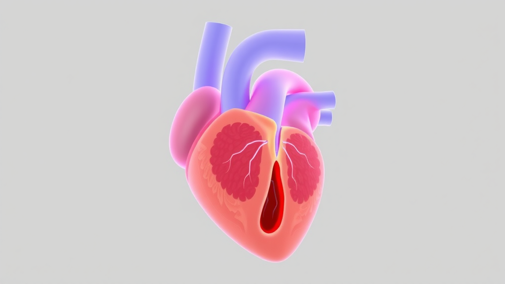
Surgeons who rebuild the mitral valve—the structure that keeps blood flowing in one direction through the left side of the heart—have long relied on proportional rules of thumb to estimate the dimensions of the leaflets they must reconstruct. A new proof-of-concept study suggests that machine learning can do better, offering patient-specific predictions of the exact anatomical measurements needed to design individualized leaflet templates. The work, published in BioMedical Engineering OnLine, connects anatomical morphometry, statistical modeling, and digital template construction into a single workflow that could one day support more precise mitral valve repair using autologous pericardium, the patient’s own tissue.
The mitral valve sits between the left atrium and the left ventricle, and its two leaflets—anterior and posterior—must open and close tens of thousands of times a day without leaking. When the valve fails, surgeons may reconstruct the leaflets using a patch of pericardium harvested from the patient. The success of such reconstruction depends on getting the geometry right: the height, width, and free-edge length of each scallop of the leaflets determine how well the repaired valve coapts and seals during systole. Conventional proportional formulas, which estimate leaflet dimensions from a few reference measurements, provide only simplified approximations that ignore the considerable anatomical variability between individuals.
To address this gap, a team of researchers from I.M. Sechenov First Moscow State Medical University (Sechenov University) in Moscow and Universiti Malaysia Sarawak analyzed clinical and morphometric data from 72 adult autopsy cases in which the mitral valve was free of structural disease. From each case, they recorded a set of patient-level characteristics and valve-level measurements, then defined eight anatomical targets that would be required for building a leaflet template: heights and free-edge lengths of the individual anterior and posterior leaflet scallops. The posterior leaflet is typically divided into three scallops—P1, P2, and P3—while the anterior leaflet is similarly segmented into A1, A2, and A3 regions, and each of these segments has its own measurable geometry.
The researchers compared eight different regression approaches in separate target-specific modeling workflows rather than forcing a single algorithm onto all eight anatomical outputs. This design reflected an important practical insight: different anatomical dimensions may depend on different combinations of inputs and may exhibit different statistical relationships with the available predictors. After systematic comparison, the final model set consisted of four ElasticNet regressors and four random forest regressors, a split that balances the interpretability and regularization of penalized linear models against the flexibility of ensemble tree-based methods.
The clinically oriented configuration used 17 patient- and valve-level inputs to estimate the eight anatomical dimensions. Predictive performance varied considerably across targets. The most accurate predictions were achieved for the heights of the P1 and P3 scallops of the posterior leaflet, where the mean absolute error was between 1.68 and 1.70 millimeters and the coefficient of determination, R-squared, reached 0.54 to 0.55. In practical terms, the models could estimate these posterior scallop heights to within roughly a millimeter and a half to two millimeters of the true anatomical value, and they explained a moderate share of the variance across individuals. Among the anterior-leaflet parameters, the A3 height showed the best performance, with a mean absolute error of 2.33 millimeters and an R-squared of 0.21, indicating that while the average error remained clinically modest, the models captured much less of the individual-to-individual variability in this region.
The least predictable target was the free-edge length of the posterior leaflet, where the mean absolute error rose to 13.32 millimeters and the R-squared dropped to just 0.02—essentially no better than predicting the population mean. This result is anatomically meaningful rather than merely a modeling failure. The free edge of the posterior leaflet is a long, scalloped, highly variable structure, and its total length appears to depend on factors not well represented among the 17 inputs available in this dataset. The finding highlights a key limitation of the proof-of-concept stage: the models are only as informative as the morphometric and clinical variables they are given, and some dimensions of the valve may require additional predictors, such as imaging-derived measurements of the annulus, chordae, or ventricular geometry.
A central strength of the study lies in its clinical orientation. Rather than stopping at statistical performance metrics, the authors incorporated the model outputs into an interactive tool for generating individualized computer-aided leaflet templates. In the envisioned workflow, a surgeon would enter a small set of patient- and valve-level measurements, the trained models would predict the eight required anatomical dimensions, and the tool would translate those predictions into digital templates sized for the individual patient. Such templates could then be used to cut and shape an autologous pericardial patch with a precision that proportional rules cannot deliver, potentially reducing intraoperative guesswork and improving the geometric fidelity of the repair.
The authors are careful to frame the work as a proof of concept rather than a clinical tool ready for the operating room. The dataset of 72 anatomical cases, while sufficient to train and compare regression models, is modest by machine-learning standards, and the models were developed on autopsy-derived measurements rather than on living patients. Before any clinical implementation, the researchers emphasize that a prospective comparison of imaging-derived and anatomical measurements is required to confirm that predictions based on clinical imaging data align with true anatomical geometry, followed by validation in independent cohorts. These steps are essential to ensure that the millimeter-level accuracies observed in the anatomical dataset translate into real-world surgical reliability.
The study also carries implications for how patient-specific modeling may evolve in structural heart surgery more broadly. Individualized prediction of valve geometry fits into a larger trend toward computational planning in cardiac surgery and interventional cardiology, where three-dimensional echocardiography, computed tomography, and cardiac magnetic resonance imaging increasingly feed patient-specific models that simulate device behavior and repair outcomes. A machine-learning layer that estimates otherwise unmeasurable anatomical parameters could complement these imaging pipelines, particularly in settings where advanced imaging is incomplete or where the relevant dimensions are not directly accessible. The Sechenov and UNIMAS team’s target-specific approach—fitting and selecting a different model for each anatomical output—offers a pragmatic template for other groups attempting to map clinical predictors onto complex three-dimensional anatomy.
For now, the message of the study is one of careful, incremental progress. The models perform well for some dimensions, notably the heights of the P1 and P3 posterior scallops, but struggle with others, especially the posterior free-edge length, and the authors do not overstate their case. What they have demonstrated is that the full chain—from anatomical measurement, through machine-learning estimation, to digital template construction—is technically feasible, ethically approved, and open to refinement. If subsequent prospective and independent validation confirms the approach, the humble autopsy dataset could become the foundation for a new generation of personalized mitral valve reconstructions, in which the leaflet a surgeon implants is not an approximation drawn from population averages but a prediction tailored to the anatomy of a single patient.
Subject of Research: Machine-learning prediction of patient-specific mitral valve anatomical parameters for reconstructive planning
Article Title: Machine-learning prediction of patient-specific mitral valve anatomical parameters for reconstructive planning
Article References: Komarov, R. N., Dydykin, S. S., Vasalatiy, I. M., Kapitonova, M., & Drakina, O. V. (2026). Machine-learning prediction of patient-specific mitral valve anatomical parameters for reconstructive planning. BioMedical Engineering OnLine. https://doi.org/10.1186/s12938-026-01624-4
Image Credits: AI Generated
DOI: 10.1186/s12938-026-01624-4
Keywords: mitral valve, machine learning, morphometry, patient-specific prediction, autologous pericardium, reconstructive planning, ElasticNet, random forest, cardiovascular models, echocardiography, predictive medicine, biomedical engineering
Cite Scienmag News
Ophelia Keating. (September 22, 2026). AI Learns to Predict Mitral Valve Anatomy for Personalized Heart Repair. Scienmag. https://scienmag.com/ai-learns-to-predict-mitral-valve-anatomy-for-personalized-heart-repair/
Ophelia Keating. "AI Learns to Predict Mitral Valve Anatomy for Personalized Heart Repair." Scienmag, 22 September 2026, https://scienmag.com/ai-learns-to-predict-mitral-valve-anatomy-for-personalized-heart-repair/. Accessed 4 October 2026.
Ophelia Keating. "AI Learns to Predict Mitral Valve Anatomy for Personalized Heart Repair." Scienmag. September 22, 2026. https://scienmag.com/ai-learns-to-predict-mitral-valve-anatomy-for-personalized-heart-repair/


