A molecular gatekeeper that decides when embryonic brain cells commit to becoming astrocytes has been identified in a new study of mouse cortical development. The protein, PCGF1, is a core component of a Polycomb repressive complex, and the research shows that its loss selectively blocks the production of astrocytes in the developing neocortex while leaving neurons largely untouched. The work, published as a Report in EMBO Reports, traces this effect to a surprising route: rather than switching astrocyte genes on directly, PCGF1 silences a gene called N4bp1, whose product destabilizes the Notch intracellular domain, the active form of a signaling molecule long known to drive astrocyte differentiation.
Astrocytes are generated from the same multipotent neural progenitor cells that produce neurons during corticogenesis, the process that builds the six-layered neocortex. Established work has shown that transcription factors such as NFIA and SOX9 initiate the astroglial program, while signaling pathways including JAK-STAT, BMP and Notch act as central regulators. Notch sustains progenitor pools during early neurogenesis and later promotes astrogliogenesis by activating glial genes such as Nfia. What remained poorly understood was how epigenetic mechanisms establish the competence of progenitors to respond to these extracellular cues. The new study addresses that gap by focusing on PRC1.1, one of six Polycomb repressive complex 1 variants defined by different PCGF subunits, which repress transcription through monoubiquitination of histone H2A at lysine 119.
To follow Pcgf1 expression over time, the researchers analyzed two published single-cell RNA sequencing atlases of the developing mouse neocortex, covering stages from embryonic day 10 to postnatal day 7. Pcgf1 transcripts were detected at low levels across multiple neural cell types, including radial glial cells, intermediate progenitors, neurons and glial populations, without strong cell type specificity. A modest increase was observed during mid-to-late embryonic stages, approximately E14.5 to E17.5. More direct measurements pointed the same way: quantitative RT-PCR of neural progenitor cells isolated at E14.5, E16.5 and E18.5 showed a progressive rise in Pcgf1 mRNA, peaking at E18.5, and immunoblotting showed increasing PCGF1 protein toward E18.5 that remained high at postnatal day 0. This timing coincides with the gliogenic window, when cortical progenitors progressively acquire astroglial characteristics.
The team then generated dorsal forebrain-specific conditional knockout mice by crossing Pcgf1-floxed animals with D6-Cre mice, whose recombination activity is largely restricted to the dorsal telencephalon. Deletion efficiency was confirmed at both mRNA and protein levels. At postnatal day 10, the knockout mice weighed less than littermate controls, although cortical area and gross morphology were preserved; the authors speculate this growth effect may arise indirectly from altered forebrain regulation of feeding behavior, metabolism or neuroendocrine function. Immunostaining revealed a transient increase in PAX6-positive apical progenitors from E18.5 in the knockout cortex, while TBR2-positive intermediate progenitors were unchanged across the stages examined. Neurosphere assays using E18.5 progenitors showed that Pcgf1-deficient cells formed significantly more and larger neurospheres than controls, indicating enhanced self-renewal potential, yet Ki67 and TUNEL staining at E16.5 and E18.5 showed no significant differences in proliferation or apoptosis.
Crucially, neuronal output was largely preserved. EdU pulse-labeling experiments, in which pregnant mice received the thymidine analog at E16.5 and offspring brains were analyzed at P3, revealed no significant difference in the proportion of EdU-positive cells that became NeuroD2-positive neurons between genotypes. Short-term labeling at E16.5 or E17.5 gave the same result. Cortical lamination, assessed with layer-specific markers, showed no significant differences in the densities or distribution of CUX1-positive layer II-IV neurons, CTIP2-positive layer V neurons or TLE4-positive layer VI neurons at P3 or P7. The oligodendroglial marker SOX10 also remained unchanged. In contrast, immunostaining at E18.5 and P3 revealed a marked reduction in GFAP-positive and ALDH1L1-positive astrocytes in the knockout cortices, and SOX9-positive and BLBP-positive astroglial progenitor populations were likewise reduced at E18.5 and P3.
The authors note an important nuance: ALDH1L1 is not restricted to differentiated astrocytes during embryonic corticogenesis but is also expressed by radial glial progenitors, and ALDH1L1-positive cells remained detectable in the ventricular zone at E18.5. The persistence of these ventricular zone cells, together with the expanded PAX6-positive progenitor pool, suggests that progenitors are maintained or expanded after Pcgf1 deletion, while the loss of GFAP-positive and parenchymal ALDH1L1-positive cells indicates impaired progression toward more differentiated astroglial states. EdU fate-mapping reinforced this interpretation: after EdU administration at E17.5 and analysis at E18.5, the proportions of EdU-positive cells co-expressing SOX9 or BLBP were reduced in knockouts, whereas the EdU-positive EGFR-positive fraction was unchanged. Because EGFR marks a broad, heterogeneous gliogenic progenitor population rather than an astrocyte-committed state, the overall gliogenic progenitor pool appears largely intact, pointing to a selective impairment of astroglial lineage progression rather than a global gliogenesis defect.
Cell culture experiments supported a cell-intrinsic requirement. When neural progenitors isolated from E18.5 knockout and control cortices were induced to differentiate under growth factor withdrawal, Pcgf1-deficient cultures showed a significantly reduced proportion of GFAP-positive astrocytes at both three and seven days, while the percentage of MAP2-positive neurons was largely unaffected. Conversely, overexpressing PCGF1 in wild-type E18.5 progenitors by electroporation significantly increased the proportion of GFAP-positive astrocytes, with no detectable changes in MAP2-positive neurons or SOX9-positive cells. To probe the underlying molecular program, the team performed RNA sequencing on FACS-purified CD133-positive progenitors from E18.5 cortices, identifying 349 upregulated and 140 downregulated genes at a log2 fold-change threshold of 1 and an adjusted P value below 0.05. Gene Ontology analysis flagged terms related to gliogenesis, glial development and glial differentiation, and independent RT-qPCR confirmed reduced expression of the glial genes S100b, Sox9, Gfap, Nfia and Nfic.
The Notch connection emerged from this screen. Expression of the canonical Notch target genes Hey1 and Hes5 was significantly reduced upon Pcgf1 deletion, whereas Notch1 transcript levels were unchanged. Immunoblotting revealed a marked reduction in the active Notch intracellular domain, accompanied by accumulation of full-length NOTCH1 and its transmembrane fragment, suggesting additional regulation at the level of receptor processing, trafficking or turnover. Among known regulators of NICD turnover, including N4bp1, Nedd4, Cbl, Dtx1, Fbxw7 and Itch, only N4bp1 was consistently upregulated at both mRNA and protein levels in Pcgf1-deficient progenitors. PCGF1 overexpression reduced endogenous N4BP1 protein in N1E-115 cells, and in HeLa cells PCGF1 overexpression decreased while PCGF1 knockdown increased endogenous N4BP1 mRNA. Because no ChIP-grade PCGF1 antibody was available, the researchers turned to published ChIP-seq data from mouse embryonic stem cells, which showed a prominent PCGF1 binding peak near the N4bp1 promoter, along with H2AK119ub and H3K27me3 enrichment that decreased following Pcgf1 loss. A luciferase reporter carrying a roughly 1.5-kilobase N4bp1 promoter fragment was repressed by ectopic PCGF1 in both N1E-115 and HeLa cells and enhanced by PCGF1 depletion in HeLa cells.
The reciprocal experiment clinched the axis. In a D6-Cre-driven N4bp1 conditional knockout, immunostaining of P3 cortices revealed a significant increase in ALDH1L1-positive, GFAP-positive and SOX9-positive cells, a phenotype opposite to that of Pcgf1 loss. The authors propose that PCGF1 operates through a double-negative mechanism: by repressing N4bp1, which encodes a negative regulator of NICD stability, PCGF1 maintains Notch signaling activity and thereby permits appropriate interpretation of developmental signals, functioning as a permissive epigenetic regulator rather than a direct fate determinant. They also place the finding in context: deletion of the core PRC1 catalytic subunit RING1B from neural progenitors impairs astrocyte differentiation, whereas loss of Pcgf2/4 increases astrocyte generation and deletion of Pcgf3/5 yields no overt change, highlighting functional diversity among PRC1 variants. The Pcgf1 phenotype is more selective than the RING1B phenotype, suggesting PRC1.1 specializes in astroglial progression rather than acting as a universal neurogenic-to-gliogenic switch.
The authors acknowledge limitations. Direct regulation of N4bp1 by PCGF1 was inferred from published ChIP-seq datasets generated in embryonic stem cells and from promoter-reporter assays rather than chromatin profiling in cortical progenitors; formal genetic epistasis between Pcgf1 and N4bp1 remains to be established, with NICD rescue or double conditional knockout models planned as future tests; the lack of suitable antibodies prevented reliable assessment of endogenous PCGF1 distribution across cortical cell populations; and the long-term persistence of, or compensation for, the astroglial defects after Pcgf1 loss remains undetermined. Subtle effects on later oligodendrocyte maturation also cannot be excluded. Even so, the study establishes the PCGF1-N4BP1-Notch axis as a candidate molecular link between Polycomb-mediated chromatin regulation and developmental signaling, expanding current models of how chromatin regulators shape neural lineage diversification by tuning progenitor sensitivity to extracellular cues.
Subject of Research: Epigenetic regulation of astrocyte development in the mouse cerebral cortex by the Polycomb protein PCGF1
Article Title: PCGF1/PRC1.1 selectively sustains astroglial lineage progression by repressing N4bp1 to stabilize Notch signaling
Article References: Yin, L., Zhou, J., Ma, Z., Wang, C., Shen, M., Hou, L., Yin, B., Qiang, B., Shu, P., & Peng, X. (2026). PCGF1/PRC1.1 selectively sustains astroglial lineage progression by repressing N4bp1 to stabilize Notch signaling. EMBO Reports. https://doi.org/10.1038/s44319-026-00983-2
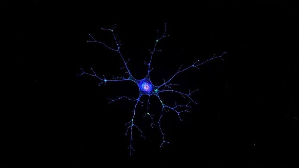
Image Credits: AI Generated
DOI: 10.1038/s44319-026-00983-2
Keywords: PCGF1, PRC1.1, Polycomb, astrocytes, Notch signaling, N4BP1, neural progenitor cells, cortical development, gliogenesis, epigenetics, conditional knockout, EMBO Reports
Cite Scienmag News
Juliet Wilcox. (October 29, 2026). Polycomb Protein PCGF1 Steers Astrocyte Development by Guarding Notch Signals. Scienmag. https://scienmag.com/polycomb-protein-pcgf1-steers-astrocyte-development-by-guarding-notch-signals/
Juliet Wilcox. "Polycomb Protein PCGF1 Steers Astrocyte Development by Guarding Notch Signals." Scienmag, 29 October 2026, https://scienmag.com/polycomb-protein-pcgf1-steers-astrocyte-development-by-guarding-notch-signals/. Accessed 30 October 2026.
Juliet Wilcox. "Polycomb Protein PCGF1 Steers Astrocyte Development by Guarding Notch Signals." Scienmag. October 29, 2026. https://scienmag.com/polycomb-protein-pcgf1-steers-astrocyte-development-by-guarding-notch-signals/