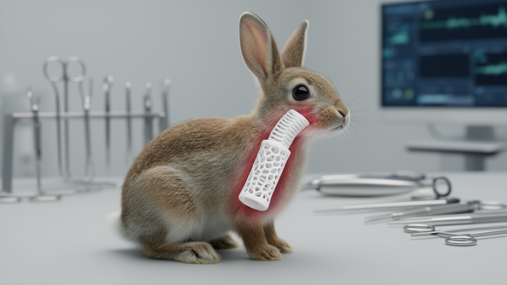
Researchers in Iran have built a tracheal patch from a 3D printer and implanted it into the necks of nine rabbits, reporting that the synthetic tube kept the animals’ airways fully open for a month. The study, published in BMC Biomedical Engineering, tested a scaffold made from a polyurethane-polyvinyl chloride composite in a model of partial tracheal wall reconstruction, and its results offer both an encouraging proof of concept and a sobering reminder of how far engineered airway substitutes still have to go.
The team, led by Ali Tadayon and Mobin Haghdel of Shiraz University of Medical Sciences, set out to address one of thoracic surgery’s most stubborn problems. When a section of the trachea must be removed because of tumors, trauma, fistulas or congenital narrowing, surgeons can only reconnect the remaining ends directly if the excised segment is short enough, no more than half the tracheal length in adults and one-third in children. Beyond that limit, the segmental blood supply of the airway makes the suture line vulnerable to ischemia and dehiscence, while postoperative inflammation and granulation tissue can trigger restenosis and impaired mucus clearance. Existing alternatives, from pericardial patches and autografts to stents and decellularized cadaveric grafts, each carry burdens of infection, rejection, donor shortage or mismatched mechanical behavior.
The Shiraz group chose a deliberately simple route. Rather than engineering a biomimetic construct seeded with cells or growth factors, they fabricated a non-biodegradable scaffold from polyurethane blended with polyvinyl chloride at a ratio of 70:30 by weight. Polyurethane contributes flexibility, elasticity and tensile strength, while PVC adds rigidity and durability. The polymers were dissolved in a dimethylformamide and tetrahydrofuran solvent system at a concentration of 18 to 22 percent, magnetically stirred for twelve hours at 40 degrees Celsius, and extruded into filament for printing.
The printing itself followed an extrusion-based fused deposition modeling process on a locally built machine, with a 200-micrometer nozzle operating at 180 degrees Celsius, a print speed of 10 millimeters per second and a 0/90-degree grid lattice infill at 50 percent density. The resulting tubes had an inner diameter of 8 millimeters and a length of 15 millimeters, dimensions matched to the adult New Zealand white rabbit trachea, with an average pore size of 250 to 400 micrometers and porosity of roughly 60 percent. Each tube was then sectioned lengthwise into a half-pipe patch covering about half of the tracheal circumference, vacuum-dried, cured, and sterilized by a combination of autoclaving and ethylene oxide gas.
Before implantation, the material showed a tensile strength of 25 plus or minus 3 megapascals, an elongation at break of 350 plus or minus 50 percent, a compressive strength of 800 plus or minus 100 kilopascals and an elastic modulus of 150 plus or minus 20 megapascals, properties the authors describe as robust enough to withstand coughing and respiratory pressures while remaining flexible enough to follow the trachea’s movements. Nine female rabbits, each three months old and weighing 2.5 to 3 kilograms, then underwent surgery in which a 15-millimeter anterior tracheal defect spanning about four rings was created and repaired with the sutured scaffold patch.
Eight of the nine animals survived the full 30-day observation period, a survival rate of 88.9 percent with a wide 95 percent confidence interval of 51.8 to 99.7 percent that reflects the small sample. The single death occurred on postoperative day 14 in an animal that had shown neck subcutaneous emphysema three days earlier; post-mortem examination found emphysema and purulent discharge around the reconstruction, but because microbiological cultures and formal airway integrity testing were not performed, the precise cause could not be determined. The authors list infection, suture-line failure or a combination as possible contributors.
In the eight survivors, bronchoscopic examination at the endpoint found complete luminal patency with no stenosis, obstruction, mucus plugging or purulent discharge, corresponding to the study’s highest patency grade. Yet the histological picture was considerably more mixed. Half of the rabbits, four of eight, showed favorable epithelialization, defined as epithelial coverage of more than half of the reconstructed luminal surface, accompanied by mild inflammation. Two animals had unfavorable epithelialization with less than 50 percent coverage and moderate inflammation, and two showed scant or absent epithelial coverage with severe inflammation. A blinded pathologist graded the sections on a three-point scale, and the inflammatory burden tracked inversely with epithelial coverage.
The explanted scaffolds told a story of mechanical resilience. After 30 days in vivo, tensile strength had slipped only slightly to 23 plus or minus 4 megapascals, elongation at break remained high at 320 plus or minus 60 percent, compressive strength fell to 750 plus or minus 120 kilopascals and elastic modulus declined to 145 plus or minus 25 megapascals. Porosity decreased from 60 plus or minus 5 percent to 55 plus or minus 8 percent, which the authors attribute to cellular infiltration and extracellular matrix deposition, while density remained unchanged at 1.21 plus or minus 0.07 grams per cubic centimeter, suggesting no substantial swelling or structural compromise.
The authors themselves are careful about what these findings do and do not show. Because the scaffold is non-degradable and rigid, the preserved airway caliber may have owed as much to mechanical support as to genuine tissue repair, and the discrepancy between clear bronchoscopy and incomplete mucosal regeneration in more than half of the animals suggests that short-term patency does not equal biological integration. With only 30 days of follow-up, delayed complications such as progressive granulation tissue, chronic inflammation and secondary stenosis could not be assessed, and the authors explicitly caution that the absence of stenosis at one month should not be read as evidence that late narrowing will not occur.
The limitations list is long and candid: no control or sham group, purely descriptive statistics, qualitative rather than quantitative airway measurements, a single pathologist whose inter-observer reliability could not be tested, no evaluation of how sterilization affected the material, no scanning electron microscopy or micro-computed tomography of the microstructure, and, notably, no in vitro cytotoxicity, extractable-leachable or ISO 10993 biocompatibility testing before the animal work. The authors frame the study as an early feasibility assessment of a cheap, locally fabricated platform rather than a regenerative construct, and they call for longer follow-up, comparator groups, serial bronchoscopy, comprehensive safety testing and bioactive modifications such as cell seeding or growth factors in future work. For now, the experiment demonstrates that a printed plastic patch can hold an airway open, not that it can rebuild one.
Subject of Research: A 3D-printed polyurethane-PVC scaffold for partial tracheal wall reconstruction tested in a rabbit model
Article Title: Development of a 3D-printed synthetic tracheal scaffold for partial tracheal wall reconstruction: an experimental animal study
Article References: Tadayon, A., Bahrani, M., Negahdari, T., Jahromi, M. A., Yousufzai, S., Moradi, S., & Haghdel, M. (2026). Development of a 3D-printed synthetic tracheal scaffold for partial tracheal wall reconstruction: an experimental animal study. BMC Biomedical Engineering, 8(1), Article 14. https://doi.org/10.1186/s42490-026-00117-8
Image Credits: AI Generated
DOI: 10.1186/s42490-026-00117-8
Keywords: 3D printing, tracheal reconstruction, tissue engineering, polyurethane, polyvinyl chloride, rabbit model, airway patency, epithelialization, biomedical engineering, preclinical study, scaffold, tracheal stenosis
Cite Scienmag News
Denise Maddox. (October 28, 2026). 3D-Printed Synthetic Tracheal Patch Keeps Rabbit Airways Open for 30 Days. Scienmag. https://scienmag.com/3d-printed-synthetic-tracheal-patch-keeps-rabbit-airways-open-for-30-days/
Denise Maddox. "3D-Printed Synthetic Tracheal Patch Keeps Rabbit Airways Open for 30 Days." Scienmag, 28 October 2026, https://scienmag.com/3d-printed-synthetic-tracheal-patch-keeps-rabbit-airways-open-for-30-days/. Accessed 28 October 2026.
Denise Maddox. "3D-Printed Synthetic Tracheal Patch Keeps Rabbit Airways Open for 30 Days." Scienmag. October 28, 2026. https://scienmag.com/3d-printed-synthetic-tracheal-patch-keeps-rabbit-airways-open-for-30-days/