A 32-year-old man who had been unable to father a child after a year of trying walked into an andrology clinic carrying a puzzle that had already defeated one imaging study. Three semen analyses confirmed azoospermia, the complete absence of sperm in his ejaculate, yet his semen volume and pH were normal, his testicles were normal in size and consistency, and both vasa deferentia could be felt on examination. His hormones were unremarkable, with a follicle-stimulating hormone level of 2.4 mIU/mL, luteinizing hormone of 1.9 mIU/mL and total testosterone of 17.5 nmol/L. Genetic testing, including cystic fibrosis mutation screening, karyotype analysis and Y chromosome microdeletion testing, was negative. A formal scrotal ultrasound performed earlier had even been reported as normal. Everything pointed away from a failure of sperm production and toward a blockage somewhere along the reproductive tract, but no study had yet shown where.
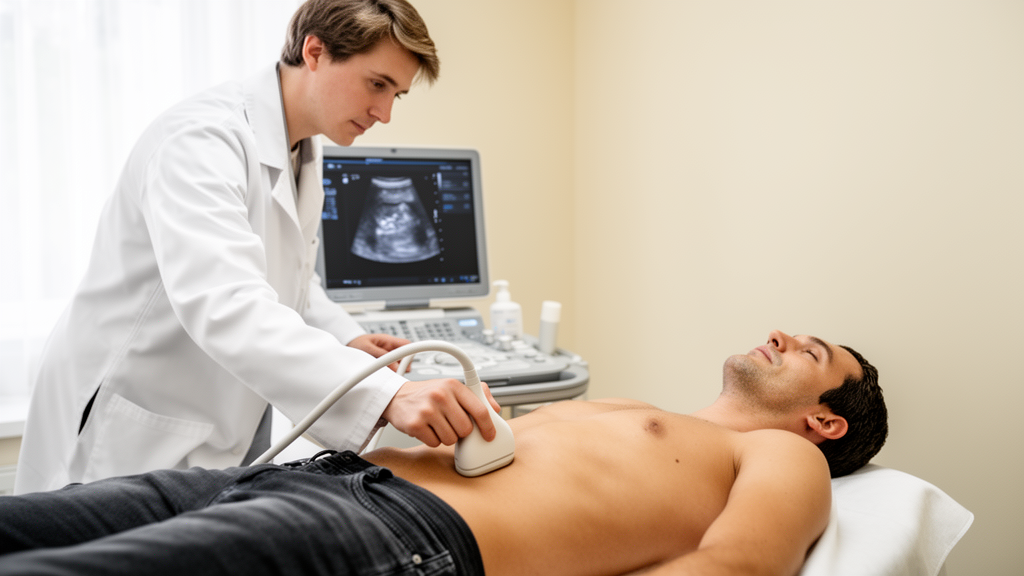
The answer came from a handheld ultrasound probe wielded not by a radiologist but by the treating andrologist, at the bedside, during that first clinic visit. Using a high-frequency linear transducer in a point-of-care ultrasonography, or POCUS, examination, the clinician found that both testes looked unremarkable, with no hypoechogenic findings. The epididymides, however, were bilaterally enlarged and studded with numerous small cysts, each under 5 millimeters, scattered within the head and tail of the structure on both sides. There was no varicocele and no hydrocele. Those sonographic features, interpreted together with the clinical history, physical examination and hormonal profile, suggested that the azoospermia was likely obstructive rather than a consequence of failed sperm production.
The case, published as a report in Basic and Clinical Andrology, matters for two reasons. Second, it spotlights a surgical history that could easily have been overlooked: as an infant, at four months of age, the patient had undergone bilateral Cohen cross-trigonal ureteral reimplantation, a pediatric operation to correct severe grade V vesicoureteral reflux, the retrograde flow of urine from the bladder back up the ureters.
Vesicoureteral reflux is a common congenital abnormality in which urine flows backward from the bladder into the ureter because of dysfunction at the ureterovesical junction. Surgical correction is highly effective and is associated with excellent long-term outcomes, and the Cohen cross-trigonal technique remains one of the most widely performed intravesical approaches. But the operation requires careful dissection near the bladder trigone, an anatomical neighborhood in which the vas deferens, the duct that carries sperm from the testis toward the urethra, lies in close proximity. The authors of the report note that while direct data on vas deferens injury during Cohen reimplantation are scarce, the pediatric and genitourinary surgery literature recognizes vas deferens injury as a risk during pelvic procedures involving the ureter or spermatic cord.
In this patient, the connection between the childhood operation and the adult diagnosis remains an association rather than a proven cause. The authors are explicit on this point: a causal relationship cannot be established from a single case, and the exact mechanism of obstruction, whether vasal injury or something else, could not be determined without invasive diagnostics that were not pursued once sperm retrieval succeeded. Detailed documentation of the original surgery’s indications and timing was not available for review, though records indicated the operation and recovery were uneventful, with no urinary tract infections or kidney problems afterward. Alternative causes of obstruction, including prior inguinal or scrotal surgery, postinfectious epididymal blockage, congenital vasal anomalies, distal seminal tract obstruction, trauma and idiopathic obstruction, were considered and not supported by the clinical picture.
What the bedside scan changed was the speed and direction of management. With the working diagnosis of obstructive azoospermia established from the POCUS findings, the patient was counseled on his options: sperm retrieval with assisted reproductive technologies, or referral for evaluation of potential reconstructive surgery. He chose to proceed directly with sperm retrieval for in vitro fertilization with intracytoplasmic sperm injection. Unilateral testicular sperm aspiration, known as TESA, yielded abundant motile sperm, a result that itself strongly supported the obstructive diagnosis, since motile sperm in the testis indicate that production is intact and the problem lies downstream. Twenty vials were cryopreserved for future use, and histopathologic examination of the biopsy specimen confirmed normal spermatogenesis, identifying spermatozoa within several of the fewer than 25 seminiferous tubules sampled.
The authors place the case in the context of their earlier research, which reported that scrotal POCUS performed by a fertility specialist or andrologist showed high diagnostic performance in distinguishing obstructive from nonobstructive azoospermia in patients with normal semen volume and pH, with a reported positive predictive value of 96.4 percent and a negative predictive value of 100 percent. Those figures come from a study of selected patients and should be read with that context in mind, but they explain why the group argues that a focused bedside examination can reduce the need for empirical exploratory surgery in appropriate cases. Obstructive azoospermia is, after all, a potentially treatable cause of male infertility, and distinguishing it from nonobstructive azoospermia directly shapes counseling and treatment. Current guidance, including AUA/ASRM recommendations, offers testicular biopsy to men with normal FSH levels and normal testicular volume when the etiology remains unclear, but the typical azoospermic man may otherwise face a long sequence of investigations: radiologist-performed scrotal ultrasound, transrectal ultrasound of the prostate and seminal vesicles, laboratory tests and sometimes diagnostic biopsy.
The report also carries a message about medical history that crosses decades and specialties. Azoospermia is found in approximately 10 to 15 percent of men undergoing fertility evaluation, and the authors argue that a seemingly distant pediatric surgical history can be pertinent when adult men present with infertility. Men with significant childhood genitourinary surgeries, such as ureteral reimplantation, may warrant early imaging evaluation of the reproductive tract, the authors suggest, even though long-term outcomes after such operations are generally positive and late complications are rare. They frame the case as an argument for multidisciplinary awareness: pediatric urologists should know that, in extremely rare circumstances, complex childhood genitourinary surgery may become relevant during a future infertility evaluation, while adult practitioners should feel confident consulting pediatric surgical records or colleagues when facing unusual infertility cases.
The authors are careful about the limits of what a single patient can teach. A case report inherently restricts generalizability, the clinical findings support only a possible association, and the mechanism of obstruction was never confirmed. They call for larger cohort studies to investigate long-term fertility outcomes in men who underwent Cohen ureteral reimplantation in infancy, with prospective tracking of fertility parameters and detailed imaging to quantify the risk of obstructive azoospermia and clarify the anatomical mechanisms involved. They also call for research into standardized POCUS protocols for fertility specialists, to support wider adoption and better diagnostic accuracy. Handheld ultrasound devices are now widely available, and studies have shown that appropriately trained non-radiologists can perform focused examinations reliably, but the evidence base in andrology remains young.
For the patient in this report, the sequence was straightforward: a bedside scan revealed multiple bilateral epididymal cysts, the childhood operation was weighed alongside the clinical picture, sperm was retrieved and frozen within the same pathway, and a diagnosis that might otherwise have required a cascade of investigations was reached efficiently. The authors describe the patient and his partner as relieved to receive a clear diagnosis and plan. Whether the childhood reimplantation truly caused the obstruction will require the population-level studies the authors propose; what the case demonstrates now is that a probe, a trained clinician and an attentive reading of an old surgical history can, in selected patients, turn a diagnostic dead end into a workable plan.
Subject of Research: Point-of-care ultrasound evaluation of obstructive azoospermia after childhood ureteral reimplantation
Article Title: Point-of-care ultrasound evaluation of obstructive azoospermia in a patient with prior bilateral Cohen ureteral reimplantation: A case report
Article References: Barak, S., Waldenberg, N., Bar, G., Barel, O., Dekalo, S., Zeadna, A., Samueli, B., & Shrem, G. (2026). Point-of-care ultrasound evaluation of obstructive azoospermia in a patient with prior bilateral Cohen ureteral reimplantation: A case report. Basic and Clinical Andrology, 36(1), Article 24. https://doi.org/10.1186/s12610-026-00321-5
Image Credits: AI Generated
DOI: 10.1186/s12610-026-00321-5
Keywords: obstructive azoospermia, point-of-care ultrasound, male infertility, ureteral reimplantation, vesicoureteral reflux, TESA, epididymal cysts, case report, andrology, sperm retrieval, Point-of-care, ultrasound
Cite Scienmag News
Ophelia Keating. (October 28, 2026). Bedside ultrasound spots blocked sperm ducts after childhood bladder surgery. Scienmag. https://scienmag.com/bedside-ultrasound-spots-blocked-sperm-ducts-after-childhood-bladder-surgery/
Ophelia Keating. "Bedside ultrasound spots blocked sperm ducts after childhood bladder surgery." Scienmag, 28 October 2026, https://scienmag.com/bedside-ultrasound-spots-blocked-sperm-ducts-after-childhood-bladder-surgery/. Accessed 28 October 2026.
Ophelia Keating. "Bedside ultrasound spots blocked sperm ducts after childhood bladder surgery." Scienmag. October 28, 2026. https://scienmag.com/bedside-ultrasound-spots-blocked-sperm-ducts-after-childhood-bladder-surgery/