


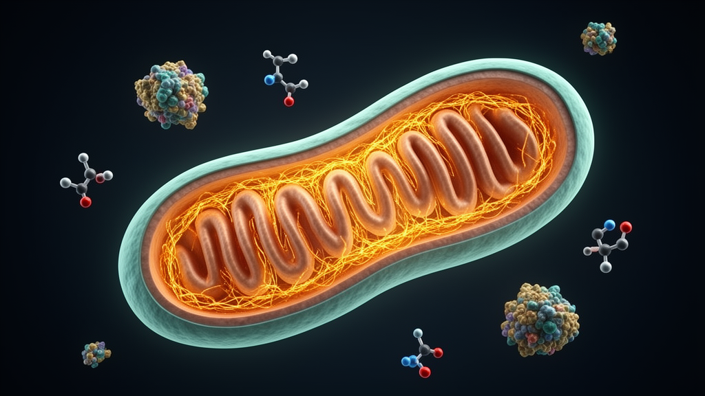
In a discovery that redraws one of the most basic diagrams in cell biology, researchers at the University of Ottawa have identified a protein that acts as a rigid molecular pillar inside mitochondria, holding the organelle’s two membranes a fixed distance apart and thereby genetically controlling the volume of the narrow compartment between them. The protein, named SPRIZ—short for SPatial Regulator of the mitochondrial Intermembrane Zone—was described in a study published in Science Advances on 14 October 2026, led by Dr. Luca Pellegrini, full professor in the Faculty of Medicine’s Department of Biochemistry, Microbiology and Immunology. The finding establishes that the intermembrane space, the thin gap sandwiched between a mitochondrion’s outer and inner membranes, is not a passive byproduct of organelle assembly but a actively regulated dimension that cells can widen or narrow to tune how their energy-producing machinery works.
Mitochondria supply most of the energy that cells need, and their ability to do so depends on a distinctive architecture: two membranes, one nested inside the other, separated by a narrow gap called the intermembrane space, or IMS. This tiny compartment helps sustain the chemistry of energy production, which makes its organization critical to how these cellular powerpacks function. The new study reports that the volume of this space is genetically controlled and adjustable. By determining where the space expands or narrows within each mitochondrion, cells can actively shape the internal architecture of their powerpacks and adjust how they operate, a level of architectural control that had not previously been demonstrated for this compartment.
To uncover the protein responsible, the team combined computer simulations of molecular movement, genetics, large-scale protein analysis and advanced imaging, including three-dimensional electron tomography. Working with collaborators at the University of Ottawa and in the United States, the researchers showed how SPRIZ bridges the two mitochondrial membranes, forming a molecular pillar that holds open the space between them. According to the article title, SPRIZ—also known by its gene name CCDC127—is a rigid tether between the outer mitochondrial membrane and the inner mitochondrial membrane that locally sets mitochondrial IMS volume, and the protein is conserved in deuterostome animals.
What makes SPRIZ distinctive, Pellegrini explains, is the way it uses a familiar molecular strategy for an unfamiliar purpose. Many membrane tethers bring membranes close enough to one another to exchange materials. SPRIZ instead appears to use that connection to maintain a defined spacing. “What makes SPRIZ so unexpected is that it uses a familiar mechanism—a tether—to do something different: it connects the two mitochondrial membranes while acting as a rigid pillar that holds them a set distance apart, so the room between them keeps a defined height,” Dr. Pellegrini explains. In other words, the same class of molecule that usually draws membranes together here props them apart, behaving less like a zipper and more like a spacer bar.
The team put this molecular spacing system to the test in laboratory experiments. Altering it substantially changed the size of the intermembrane space. Like adjusting the height of a ceiling, changing these molecular supports reshaped the room within. The findings show that cells carry genetic instructions for setting the dimensions of this essential space, giving them control over the internal layout of their powerpacks. The researchers also demonstrated that the space can be tailored: by engineering a longer version of the molecular pillar, they pushed the membranes farther apart and measured how the expanded intermembrane space affected molecular access and calcium uptake. That ability to tune the dimensions of the IMS gives researchers an experimental tool for testing whether different functions within mitochondria require different amounts of space.
Even a tiny change in the room between the membranes can alter what happens inside it. When the team expanded the IMS, molecules gained greater access to the compartment, while mitochondrial calcium uptake changed—evidence that the compartment’s dimensions help control how signals reach the machinery within these cellular powerpacks. “A change of only a few nanometres in the height of that room is enough to slow the calcium that must cross it to reach the reactor,” Dr. Pellegrini says. Because calcium handling is one of the central signaling and metabolic functions of mitochondria, the result links a nanometre-scale structural measurement directly to a measurable physiological readout.
Experiments in mice revealed how precisely SPRIZ acts. Removing the gene altered the spacing between the mitochondrial membranes while leaving the mitochondria’s overall structure intact. The mice remained viable, which allowed the team to distinguish a targeted change in the intermembrane space from widespread damage to the powerpacks themselves. That separation matters for interpreting the results: rather than a general breakdown of mitochondrial architecture, the animals displayed a specific, isolated change in the height of the compartment, confirming that SPRIZ’s role is to set that dimension rather than to hold the organelle together in a broader sense.
The study also places the protein in an evolutionary frame. The team found evidence of the same molecular pillar architecture in humans and in every other vertebrate species examined. Their evolutionary analysis places the emergence of SPRIZ around the origin of vertebrates, roughly 500 million years ago, and found it absent from plants, fungi—including yeast—and invertebrates such as sponges, worms and insects. For Pellegrini, this opens a broader perspective on how cellular powerpacks evolved to meet the changing demands of animal life. As animals diversified in body size, tissues and organs, their mitochondria had to support increasingly varied tasks while adapting to fluctuations in nutrient supply and energy needs. The arrival of SPRIZ may have given vertebrate cells an additional way to meet those demands: tailoring the dimensions of the intermembrane space to the work each cell performs. The discovery thus sits within a larger evolutionary story in which mitochondria adapted not only through changes to their molecular machinery, but also through control over the space in which that machinery works.
The discovery brought together expertise in mitochondrial metabolism, physiology and evolution. At the uOttawa Faculty of Medicine, collaborators included Drs. Mary-Ellen Harper, Eric Lee, Morgan Fullerton and Yan Burelle. The team also benefited from the contributions of two prominent U.S. scientists: mitochondrial biologist Dr. György Hajnóczky and evolutionary biologist Dr. Eugene Koonin. “That scientists of this standing chose to join the effort speaks to the importance of the questions this discovery opens up,” Dr. Pellegrini says. At the heart of the effort were his students: Raman Shulinski, the study’s first author, a graduate student, and Roman Loban and Victoria Caminiti, two undergraduates who helped carry the work forward. “For young scientists at the start of their careers to play such a central role in a discovery like this shows what our students can accomplish at the University of Ottawa,” Dr. Pellegrini says.
Beyond the laboratory, the identification of a genetically controlled spacing system inside mitochondria opens a new path to understanding how disruptions in this delicate spacing may contribute to diseases in which mitochondria fail to meet cells’ energy needs, or in which damaged powerpacks are not properly maintained or cleared away, as occurs in Parkinson’s disease. The study, published in Science Advances under the DOI 10.1126/sciadv.aef352, turns a question about the smallest dimensions of mitochondria into a finding with implications across cell biology. Dr. Pellegrini’s lab is slated to move into the University of Ottawa’s Advanced Medical Research Centre when it opens in 2027; the AMRC, the biggest investment in uOttawa history, will feature 350,000 square feet of advanced research space with a dozen state-of-the-art core facilities not found anywhere else in the region, providing the infrastructure for the next phase of work on how the height of the room inside our cells’ powerpacks shapes their function.
Subject of Research: Identification of the SPRIZ protein as a rigid mitochondrial membrane tether that genetically controls intermembrane space volume
Article Title: University of Ottawa scientists uncover the molecular pillars that shape our cells’ powerpacks
Article References: University of Ottawa scientists uncover the molecular pillars that shape our cells’ powerpacks. Original publication (Bibliographic details unavailable.)
Image Credits: AI Generated
DOI: Not provided
Keywords: mitochondria, SPRIZ, CCDC127, intermembrane space, membrane tether, calcium uptake, electron tomography, vertebrate evolution, University of Ottawa, Science Advances, Parkinson's disease, cell biology
Cite Scienmag News
Drew Townsend. (October 24, 2026). Newly Found Protein SPRIZ Sets the Space Inside Mitochondria. Scienmag. https://scienmag.com/newly-found-protein-spriz-sets-the-space-inside-mitochondria/
Drew Townsend. "Newly Found Protein SPRIZ Sets the Space Inside Mitochondria." Scienmag, 24 October 2026, https://scienmag.com/newly-found-protein-spriz-sets-the-space-inside-mitochondria/. Accessed 24 October 2026.
Drew Townsend. "Newly Found Protein SPRIZ Sets the Space Inside Mitochondria." Scienmag. October 24, 2026. https://scienmag.com/newly-found-protein-spriz-sets-the-space-inside-mitochondria/


