


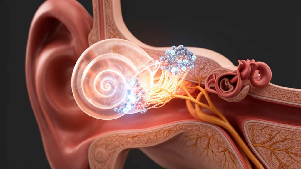
A functional study of one Han Chinese family found that a heterozygous complex insertion-deletion in exon 2 of P2RX2 disrupted pre-mRNA splicing, generated two frameshifted transcripts containing premature termination codons and markedly reduced P2X2 output in cell-based assays. The findings support a loss-of-function mechanism for the variant in autosomal dominant nonsyndromic hearing loss 41, or DFNA41, and extend the evidence for splice-disrupting alleles in this gene.
The investigators evaluated four members of a two-generation family recruited through a school for deaf children: an affected proband and three reportedly unaffected first-degree relatives. The proband passed newborn hearing screening at three days old, but subsequent testing at 19 months found no reproducible auditory brainstem responses at stimulus levels up to 97 dB nHL in either ear. Distortion-product otoacoustic emissions had signal-to-noise ratios below 6 dB at most tested frequencies bilaterally. Evaluation at age six confirmed persistent profound impairment, with click-evoked thresholds above 109 dB nHL and severe-to-profound sensorineural hearing loss across all frequencies on unaided pure-tone audiometry.
Targeted sequencing of a 406-gene hereditary hearing-loss panel identified the heterozygous P2RX2 variant NM_174873:c.278_288delinsACGTGAAGCCCCC. High-depth validation yielded mean coverage above 1,000 times, and the variant was absent from population databases including gnomAD. Segregation analysis showed that the proband inherited the P2RX2 allele from the father. The investigators classified it as pathogenic under the American College of Medical Genetics and Genomics and Association for Molecular Pathology framework using PVS1, PS3 and PM2_Supporting evidence.
The proband also carried the recurrent pathogenic GJB2 frameshift NM_004004:c.235delC, or p.Leu79Cysfs*3, inherited from the mother. The mother and younger sister carried this GJB2 allele but were reported to be unaffected. Because heterozygous GJB2 carriers are typically asymptomatic, the pedigree alone cannot establish that the GJB2 variant caused or modified the proband’s hearing loss. The authors instead treated a possible interaction between the two genes as a hypothesis requiring independent validation.
To test the splice defect, the researchers constructed pcMINI-C minigene reporters containing P2RX2 exon 2, flanking intronic sequence and downstream exon 3. The wild-type reporter yielded one RT-PCR product in HEK293T and HeLa cells, whereas the mutant reporter yielded two aberrant products in both cell lines. Sequencing resolved a 19-base-pair deletion in the first transcript and a 34-base-pair deletion in the second. Both were out-of-frame alterations that introduced premature termination codons, rather than in-frame deletions.
The reconstructed isoforms were designated mut1, corresponding to c.278_309delinsACGTGAAGCCCCCGA and predicted p.Trp93Tyrfs*173, and mut2, corresponding to c.276_309del and predicted p.Trp93Glyfs*26. Three computational tools supported the minigene result. SpliceAI assigned a score of 0.36 to loss of the canonical donor and 0.97 to gain of a cryptic donor. RDDCSC predicted a 36-base-pair deletion and an event score that increased from 0.004 in the wild type to 0.9623 in the mutant, with p=0.006. BDGP identified cryptic donor gains with scores of 0.49 and 0.68.
A SpliceVault search documented use of a cryptic donor site 19 base pairs upstream of the canonical site in 116 GTEx and 217 SRA reference samples, where it represented a rare mis-splicing event. The investigators then expressed each reconstructed isoform separately from intronless cDNA vectors in HEK293T cells, using biological triplicates. Relative to wild-type P2RX2 mRNA, mut1 abundance was lower by approximately 48.3% (p<0.05), while mut2 abundance was lower by 69.4% (p<0.01).
Western blotting detected the expected wild-type protein at about 47 kDa and truncated products at approximately 32 kDa for mut1 and 15 kDa for mut2. Densitometry normalized to GAPDH showed respective protein reductions of 92.7% and 61.0% compared with wild type, both at p<0.0001. These cell-based measurements connect the altered splicing with substantially reduced P2X2 output. Murine cochlear single-cell transcriptomic data showed P2RX2 expression mainly in sensory hair cells and spiral ganglion neurons, whereas GJB2 was enriched in supporting-cell populations, including the inner sulcus and Deiters’ cells. Computational enrichment and network analyses identified shared functional themes and nominated RYR3 as a possible connector, but these mouse-data associations did not demonstrate a digenic mechanism in the family.
The proband’s profound hearing loss contrasted with the father’s milder reported phenotype despite their shared P2RX2 allele, which the authors interpreted as consistent with variable expressivity and incomplete penetrance in P2RX2-linked DFNA41. The study was limited by its single affected proband carrying two variants, the absence of quantitative audiometry and a noise-exposure history for the father, and unavailable family-wide longitudinal audiologic tracking; the proband itself was assessed at 19 months and again at age six. Unmeasured environmental exposures and other genetic modifiers could not be excluded. The minigene and heterologous-cell experiments established the splice defect and reduced receptor output in the tested systems but did not fully reproduce the native cochlear environment. The reported evidence therefore supports the P2RX2 loss-of-function effect, while the cause of the marked intrafamilial variation remains unresolved.
Subject of Research: Functional characterization of a P2RX2 exon-2 splice-disrupting indel in a one-family DFNA41 study
Article Title: A P2RX2 exon-2 complex indel disrupts splicing and reduces P2X2 output: implications for DFNA41 and variable expressivity
Article References: Tao, Y., Song, W., Wang, L., Han, T., Han, D., Hao, L., Zhao, C., Ping, J., & Li, X. (2026). A P2RX2 exon-2 complex indel disrupts splicing and reduces P2X2 output: implications for DFNA41 and variable expressivity. Frontiers in Genetics, 17, Article 1934538. https://doi.org/10.3389/fgene.2026.1934538
Image Credits: AI Generated
DOI: 10.3389/fgene.2026.1934538
Keywords: P2RX2, DFNA41, hearing loss, complex indel, aberrant splicing, P2X2 receptor, loss of function, minigene assay, GJB2, variable expressivity, exon-2, complex
Cite Scienmag News
Drew Townsend. (October 21, 2026). P2RX2 exon-2 indel disrupts splicing and reduces receptor output in one DFNA41 family. Scienmag. https://scienmag.com/p2rx2-exon-2-indel-disrupts-splicing-and-reduces-receptor-output-in-one-dfna41-family/
Drew Townsend. "P2RX2 exon-2 indel disrupts splicing and reduces receptor output in one DFNA41 family." Scienmag, 21 October 2026, https://scienmag.com/p2rx2-exon-2-indel-disrupts-splicing-and-reduces-receptor-output-in-one-dfna41-family/. Accessed 21 October 2026.
Drew Townsend. "P2RX2 exon-2 indel disrupts splicing and reduces receptor output in one DFNA41 family." Scienmag. October 21, 2026. https://scienmag.com/p2rx2-exon-2-indel-disrupts-splicing-and-reduces-receptor-output-in-one-dfna41-family/


