


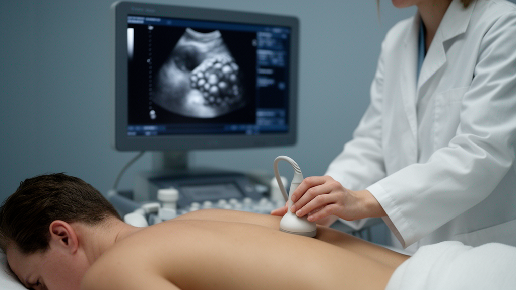
For decades, doctors assessing kidney health in people with high blood pressure have relied almost entirely on numbers: an estimated glomerular filtration rate calculated from a blood sample, and a dipstick test for protein in the urine. Both are functional measures, and both have a well-known blind spot. By the time they turn abnormal, substantial structural damage may already have taken root inside the kidneys. Now, a research team in China reports that the answer may be visible on the surface of the organ itself, detectable with the same ultrasound machines already humming away in health screening centers around the world.
The new study, published in Frontiers in Endocrinology, set out to translate a concept that had previously lived only in the world of contrast-enhanced CT scanning. Investigators at the Mayo Clinic had shown years earlier that microscopic nephron hypertrophy and nephrosclerosis, the hallmarks of hypertensive kidney injury, manifest macroscopically as increased roughness and nodular contour change on the renal surface. Subsequent CT-based studies linked this renal surface nodularity, or RSN, with arterial hypertension, early renal injury, adverse vascular events, declining kidney function, and progression of renal dysfunction. But CT requires ionizing radiation and iodinated contrast, making it unsuitable for routine screening. Ultrasound, by contrast, is widely accessible, radiation-free, and cost-effective, though conventional grayscale parameters such as renal length, cortical thickness, and cortical echogenicity have shown limited sensitivity for detecting early structural change.
The team, led by Xin Wang and Yue Chen of Wuxi Second Hospital of Traditional Chinese Medicine and Qingjian Xu of the Taihu Lake Cadre Sanatorium of Jiangsu Province, enrolled 871 hypertensive adults who underwent renal ultrasound during routine health screening between October 2025 and March 2026. The cohort was drawn from a screening population rather than a nephrology clinic, and patients with myocardial infarction or atrial fibrillation were excluded because those conditions can cause focal cortical scarring or independently affect renal hemodynamics. The median age was 58 years, 83.2 percent were men, and 21.8 percent had diabetes. All participants had stored standardized coronal-plane ultrasound images of the kidneys acquired on a GE LOGIQ E11 system with a convex array transducer operating at 4.5 megahertz.
The technical standardization was deliberate. For each kidney, coronal-plane scanning through the renal hilum was performed, and a single representative static frame was selected for grading using predefined criteria: a hilar section showing maximal renal length, a complete and clearly delineated capsule, no appreciable respiratory-motion or reverberation artifact, and clear differentiation between parenchyma and sinus. Subjects breathed quietly without breath-holding, and uniform light transducer pressure was applied to avoid deforming the contour. Imaging parameters were fixed across examinations, with a dynamic range of 60 to 80 decibels, gain adjusted to display the capsule clearly, a single near-field focal zone at the capsule, tissue harmonic and spatial compound imaging enabled, and depth adjusted to body habitus between 10 and 15 centimeters.
Grading followed a three-point scale adapted from the previously validated CT-based system. Grade 0 described a smooth renal capsule with intact continuity. Grade 1, termed mild and undulating, described a wave-like contour or focal indentations with a depth less than one-third of the cortical thickness. Grade 2, severe and serrated, described a nodular contour with indentation depth greater than one-third of the cortical thickness, or accompanying cortical thinning. A sonographer with more than ten years of experience, blinded to laboratory results, graded the stored images. For analysis, the maximum grade of the two kidneys was used as the patient-level score, chosen a priori because hypertensive nephrosclerosis may be asymmetric, though bilateral concordance proved high, with agreement in 88.2 percent of cases and a weighted kappa of 0.888.
Reproducibility, essential for any clinical adoption, was assessed in a random subset of 80 patients. A second sonographer independently evaluated the images while blinded to the first reader’s results, and the first reader re-evaluated the same subset after at least four weeks. Interobserver agreement for the patient-level RSN score yielded a weighted kappa of 0.798, with 80 percent agreement, indicating substantial agreement. Intraobserver agreement showed a weighted kappa of 0.888, with 87.5 percent agreement, indicating almost perfect agreement. For individual kidney assessment, both interobserver and intraobserver kappa values exceeded 0.83. Cortical echo intensity, graded from lower than liver or spleen to higher than liver or spleen, also demonstrated substantial-to-almost-perfect reproducibility.
The prevalence findings were striking. RSN was detected in 410 of 871 patients, or 47.1 percent: 226 patients, 25.9 percent, had grade 1, and 184, 21.1 percent, had grade 2. Higher grades tracked with older age, male sex, higher hypertension grades, and longer hypertension duration. Median eGFR declined progressively across grades, from 98.9 to 92.7 to 79.9 milliliters per minute per 1.73 square meters. The proportion with eGFR below 60 rose from 0.7 percent in grade 0 to 22.8 percent in grade 2, and proteinuria increased from 4.8 percent to 12.5 percent. In multivariable logistic regression, four factors remained independently associated with RSN: hypertension grade, with an odds ratio of 2.40; hypertension duration, 1.62; cortical echo intensity, 1.63; and lower eGFR, 0.625 per 10-unit increase. The model showed good discrimination, with an area under the curve of 0.815, well calibrated on bootstrap internal validation with an optimism-corrected AUC of 0.806.
The association with renal damage, defined as eGFR below 60 or proteinuria of at least 1-plus, showed a clear dose-response pattern. Renal damage was present in 5.2 percent of grade 0 patients, 8.0 percent of grade 1, and 27.2 percent of grade 2, a more than fivefold increase. Grade 2 RSN remained strongly associated after adjustment for age, sex, hypertension grade, and diabetes, with an odds ratio of 8.77, and persisted after further adjustment for hypertension duration and cortical echo intensity, at 7.66. When the team additionally adjusted for eGFR as a continuous variable, the association was markedly attenuated but remained significant, at 2.88. In a sensitivity analysis restricted to the non-eGFR components of the endpoint and adjusted for continuous eGFR, the association was no longer significant, at 1.55. The authors interpret this honestly: RSN is not a marker independent of eGFR, but a morphological correlate of hypertensive renal damage that is directly visible on routine ultrasound. Coexisting diabetes conferred additional risk, with an odds ratio of 2.04, consistent with known synergistic mechanisms of renal injury.
Perhaps the most provocative finding emerged among the 779 patients whose eGFR and urinalysis remained within normal limits. Within this apparently healthy subgroup, 134 patients, 17.2 percent, already exhibited grade 2 RSN, and eGFR declined progressively across RSN grades, with medians of 98.8, 93.1, and 87.4. Conversely, 134 of the 184 patients with grade 2 RSN, or 72.8 percent, did not meet the threshold definition of renal damage. This pattern supports the hypothesis that renal surface remodeling accompanies subclinical reductions in renal function before conventional diagnostic thresholds are crossed, suggesting that structural change may be appreciable during routine screening ultrasound even when functional indices look reassuring. The concept of quantifying organ surface nodularity has already been translated from CT to ultrasound in hepatology for liver fibrosis assessment, and adding RSN grade to cortical echo intensity improved discrimination for renal damage, with the area under the curve rising from 0.646 to 0.711, indicating the two features carry partly complementary information.
The study has limitations the authors acknowledge candidly. The cross-sectional design precludes causal inference, and temporal precedence must be tested in longitudinal cohorts. Because both readers assessed identical stored images, only interpretive rather than acquisition-level reproducibility was evaluated. Renal damage was defined using a modified MDRD equation rather than the CKD-EPI 2021 equation and dipstick proteinuria, and cystatin C and quantitative albuminuria were not available. Several covariates, including smoking and antihypertensive therapy, were not recorded, and there was no normotensive control group. Cross-modality validation against CT-based RSN or histopathology was not performed and remains the necessary next translational step. Still, the proof of concept stands: ultrasound can detect and grade renal surface nodularity with reproducible reliability, and its grade shows a graded association with renal damage in hypertensive adults. For millions of people whose blood tests look normal while their kidneys quietly bear the structural scars of chronic hypertension, a bumpy outline on a routine scan may be the first visible warning that something is changing beneath the surface.
Subject of Research: Ultrasound-detected renal surface nodularity and renal damage in hypertensive adults undergoing routine health screening: a cross-sectional study
Article Title: Ultrasound-detected renal surface nodularity and renal damage in hypertensive adults undergoing routine health screening: a cross-sectional study
Article References: Wang, X., Chen, Y., Zhu, H., & Xu, Q. (2026). Ultrasound-detected renal surface nodularity and renal damage in hypertensive adults undergoing routine health screening: a cross-sectional study. Frontiers in Endocrinology, 17, Article 1934293. https://doi.org/10.3389/fendo.2026.1934293
Image Credits: AI Generated
DOI: 10.3389/fendo.2026.1934293
Keywords: Ultrasound-detected, renal, surface, nodularity, damage, hypertensive, adults, undergoing, routine, health, screening, cross-sectional
Cite Scienmag News
Jerry Hayes. (October 19, 2026). Bumpy Kidneys on Ultrasound Reveal Hidden Damage in Adults With High Blood Pressure. Scienmag. https://scienmag.com/bumpy-kidneys-on-ultrasound-reveal-hidden-damage-in-adults-with-high-blood-pressure/
Jerry Hayes. "Bumpy Kidneys on Ultrasound Reveal Hidden Damage in Adults With High Blood Pressure." Scienmag, 19 October 2026, https://scienmag.com/bumpy-kidneys-on-ultrasound-reveal-hidden-damage-in-adults-with-high-blood-pressure/. Accessed 19 October 2026.
Jerry Hayes. "Bumpy Kidneys on Ultrasound Reveal Hidden Damage in Adults With High Blood Pressure." Scienmag. October 19, 2026. https://scienmag.com/bumpy-kidneys-on-ultrasound-reveal-hidden-damage-in-adults-with-high-blood-pressure/


