


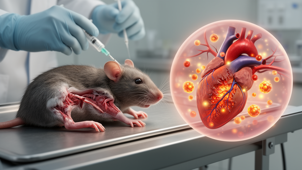
When blood flow returns to tissue after a period of ischemia, the surge of oxygen and nutrients that saves cells can also ignite a destructive inflammatory cascade. In a bilateral hind limb ischemia/reperfusion (I/R) injury model in rats, researchers tested whether the selective receptor-interacting serine/threonine-protein kinase 1 (RIPK1) inhibitor necrostatin-1s (Nec-1s) could blunt the cardiovascular collapse and inflammatory damage that follow such an insult. The findings, published in Pharmacia, suggest that blocking RIPK1 activity prevents both the hypotensive and tachycardic responses to I/R and the widespread molecular changes that drive injury in distant organs.
Necroptosis is a lytic, caspase-independent form of regulated cell death that depends on RIPK3 and mixed lineage kinase domain-like pseudokinase (MLKL) and is initiated by RIPK1. Unlike apoptosis, necroptosis ruptures the plasma membrane, causing cellular swelling, fragmentation, and release of cytoplasmic contents including damage-associated molecular patterns such as high-mobility group box 1 (HMGB1). Once outside the cell, HMGB1 acts like a cytokine, fueling inflammatory diseases. The RIPK1/RIPK3/MLKL necrosome also activates transcription factors such as nuclear factor-κB (NF-κB), increasing production of pro-inflammatory and vasodilator mediators that contribute to cardiovascular, renal, hepatic, and neurodegenerative diseases.
Ischemia develops when blood flow to tissues is insufficient, leading to oxygen deficiency, irreversible damage, and cell death as duration increases. Reperfusion, while necessary to restore oxygen and nutrients, can paradoxically exacerbate existing cellular damage and cause new injury through formation of reactive nitrogen species, reactive oxygen species, and eicosanoids via enzymes such as xanthine oxidase, inducible nitric oxide synthase (iNOS), NADPH oxidase, and cyclooxygenase-2 (COX-2). These mediators circulate and injure distant organs, producing systemic inflammation, endothelial dysfunction, and ultimately multiple organ failure (MOF). Lower-extremity I/R, which occurs in peripheral arterial disease, traumatic arterial injury, and abdominal aortic aneurysm, can damage the heart, kidney, lung, liver, and brain.
To model this, researchers applied rubber tourniquets to both hind limbs of male Wistar rats at the level of the greater trochanter under ketamine and xylazine anesthesia, inducing 3 hours of ischemia followed by 24 hours of reperfusion. Nec-1s was injected intraperitoneally 1 hour before reperfusion at doses of 1, 2, or 3 mg/kg. Mean arterial pressure (MAP) and heart rate (HR) were recorded noninvasively using a volume-pressure tail-cuff method. After 24 hours, blood was drawn from the caudal vena cava and the heart, kidney, lung, liver, and brain were isolated for molecular and histopathological analysis.
The results showed that MAP values decreased by 25 and 22 mmHg at 3 hours after ischemia and 24 hours after reperfusion, respectively, while HR values increased by 116 and 74 beats per minute over the same intervals. Nec-1s at 1, 2, or 3 mg/kg reversed the drop in MAP, but only the 3 mg/kg dose prevented the I/R-induced rise in HR. Because 3 mg/kg was the lowest effective dose that prevented both hypotension and tachycardia without mortality, it was selected for ex vivo studies.
Immunoblotting revealed that I/R increased the expression and phosphorylation of RIPK1, RIPK3, and MLKL in all five tissues examined, indicating activation of the necroptotic necrosome. HMGB1 protein expression also rose in the heart, kidney, lung, liver, and brain after I/R. Nec-1s prevented these I/R-induced increases in necrosome components and HMGB1, consistent with its role as a selective RIPK1 inhibitor that suppresses necroptotic signaling and the release of pro-inflammatory alarmins.
The study further demonstrated that I/R activated the IKKα/β/γ/IκB-α/NF-κB pathway in all five tissues, increasing phosphorylated IKKα, IKKβ, IKKγ, IκB-α, and NF-κB p65 while decreasing unphosphorylated IκB-α. Downstream, iNOS and COX-2 expression increased, as did serum and tissue nitrite levels (an indicator of iNOS-mediated NO formation) and prostaglandin I2 (PGI2) levels (an indicator of COX-2-mediated vasodilatory prostaglandin formation). Interleukin-1β expression also rose. Nec-1s prevented all of these I/R-induced molecular changes, showing that RIPK1 inhibition suppresses the transcriptional programs that generate vasodilator and pro-inflammatory mediators.
Histopathological examination using hematoxylin and eosin staining revealed mild to moderate injury-related changes in all five tissues after I/R, including inflammatory cell infiltration, edema, congestion, hemorrhage, hyperemia, necrosis, and apoptosis. Nec-1s reduced the I/R-induced increase in histopathological scores in tissues other than the liver, but the effect was statistically significant only in the heart. This suggests that while Nec-1s broadly suppresses necroptotic and inflammatory signaling at the molecular level, its ability to reverse structural tissue damage varies by organ and may require longer treatment or different dosing strategies.
The findings indicate that Nec-1s, a systemically administered drug candidate, may be beneficial in managing diseases associated with hypotension, arrhythmia, inflammation, and necroptosis caused by increased production of vasodilator mediators such as NO and PGI2 and pro-inflammatory cytokines such as HMGB1 and IL-1β. Because lower-extremity I/R injury can lead to death through MOF, a selective RIPK1 inhibitor that prevents cardiovascular deterioration and inflammatory tissue damage could represent an important therapeutic advance for patients at risk of reperfusion-related systemic collapse.
Subject of Research: Effects of RIPK1 inhibitor necrostatin-1s on cardiovascular responses and inflammatory damage in a bilateral hind limb I/R injury model in rats
Article Title: Effects of RIPK1 inhibitor necrostatin-1s on cardiovascular responses and inflammatory damage in a bilateral hind limb I/R injury model in rats
Article References: Ikiz, E., Ahmed-Reda Elosman, M., Senol, S. P., Kara, T., & Tunctan, B. (2026). Effects of RIPK1 inhibitor necrostatin-1s on cardiovascular responses and inflammatory damage in a bilateral hind limb I/R injury model in rats. Pharmacia, 73, Article e206332. https://doi.org/10.3897/pharmacia.73.e206332
Image Credits: AI Generated
DOI: 10.3897/pharmacia.73.e206332
Keywords: Effects, RIPK1, inhibitor, necrostatin-1s, cardiovascular, responses, inflammatory, damage, bilateral, hind, limb, injury
Cite Scienmag News
Ophelia Keating. (October 18, 2026). Effects of RIPK1 inhibitor necrostatin-1s on cardiovascular responses and inflammatory damage in a bilateral hind limb I/R injury model in rats. Scienmag. https://scienmag.com/effects-of-ripk1-inhibitor-necrostatin-1s-on-cardiovascular-responses-and-inflammatory-damage-in-a-bilateral-hind-limb-i-r-injury-model-in-rats/
Ophelia Keating. "Effects of RIPK1 inhibitor necrostatin-1s on cardiovascular responses and inflammatory damage in a bilateral hind limb I/R injury model in rats." Scienmag, 18 October 2026, https://scienmag.com/effects-of-ripk1-inhibitor-necrostatin-1s-on-cardiovascular-responses-and-inflammatory-damage-in-a-bilateral-hind-limb-i-r-injury-model-in-rats/. Accessed 18 October 2026.
Ophelia Keating. "Effects of RIPK1 inhibitor necrostatin-1s on cardiovascular responses and inflammatory damage in a bilateral hind limb I/R injury model in rats." Scienmag. October 18, 2026. https://scienmag.com/effects-of-ripk1-inhibitor-necrostatin-1s-on-cardiovascular-responses-and-inflammatory-damage-in-a-bilateral-hind-limb-i-r-injury-model-in-rats/


