


Breast cancer kills through its diversity. A single tumor is not one disease but a mosaic of subclones, each carrying different mutations, different receptor states, and different sensitivities to chemotherapy. This heterogeneity explains why roughly 60 percent of patients with breast cancer experience progression or recurrence despite biomarker-guided treatment, and why the field of precision oncology has spent a decade searching for laboratory models that can faithfully mirror an individual patient’s tumor before a single drug is administered. Now, a team of researchers at Prince of Songkla University in Thailand has reported a platform that may bring that goal within the clinical treatment-planning window itself: patient-derived microtumors, or PDMs, grown directly from core needle biopsies as small as a few cubic millimeters of tissue, and validated against the actual chemotherapy responses of the patients whose tumors seeded them.
The work, published in iScience, addresses a stubborn bottleneck in personalized cancer modeling. Patient-derived organoids, three-dimensional cultures grown from dissociated tumor cells, have emerged as powerful predictive models over the last several years, capable of capturing the genetic profiles and drug responses of individual tumors. But conventional organoid protocols demand substantial tissue, expensive niche-factor cocktails containing agents such as Neuregulin 1, R-spondin, FGF7/10, Noggin, and the ROCK inhibitor Y-27632, and long-term passaging that can take weeks to months. More critically, a recent study cited by the Thai team found that in an early-stage needle-biopsy setting, more than 85 percent of breast cancer organoid cultures were overtaken by normal epithelial cells, with tumor-specific alterations falling below detection within approximately two to three weeks. The tumor compartment, in other words, simply loses the competition for culture space.
The Thai researchers’ solution is mechanistically different from the organoid paradigm. Rather than fully dissociating tumor tissue into single cells that must self-organize and expand through serial passaging, the team maintained intact or non-fully dissociated tumor fragments throughout the culture period. This fragment-based approach, which the authors designate as the microtumor or ex vivo tumor fragment model, preserves native tumor architecture while shortening generation time. It also largely excludes the normal single-cell proportion during isolation, avoiding the clonal competition between normal and malignant epithelial populations that plagues dissociated organoid cultures. The distinction matters: across the short assay window, sample identity was maintained, as demonstrated by short tandem repeat profiling showing 100 percent concordance between microtumors and matched tissues in most cases, together with retention of tumor-associated BRCA1 copy-number alterations.
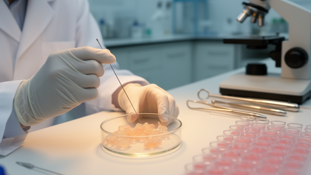
The technical workflow is elegantly simple. Core needle biopsies and small tissue specimens were minced into pieces of 0.5 to 1 cubic millimeter in a cold working medium. During this mincing step, the team developed a simple observational technique: under an inverted light microscope, the release of tumor fragments and cell clusters into the medium could be used to estimate the chance of successful microtumor derivation. This phenomenon was detected in most successful cases but was absent in failure cases, correlating with histological features visible on hematoxylin and eosin staining. Tissues that successfully yielded microtumors exhibited a higher number of cancer epithelial cells with distinct nuclei and cytoplasm, while failure cases had few cells with unclear morphology.
Following mincing, the team collected tumor fragments from two parallel isolation pathways. Mechanical isolation, achieved by gravity separation and shearing with a wide-bore pipette tip, recovered fragments from the supernatant. Enzymatic digestion of the remaining tissue sediment, using Collagenase type 2 for 15 to 30 minutes in a shaking incubator, recovered additional fragments, including smaller ones. This dual-stage fragment recovery strategy was designed to maximize the utilization of limited tumor material, which is particularly relevant for core needle biopsies. The yields varied between the two methods depending on tissue specimens, with mechanical isolation producing higher fragment or cell density in about half of the successful cases and enzymatic isolation higher in others. Because of this variability, fragments from both pathways were cultured separately to avoid yield loss.
The culture medium itself was optimized from a previously published recipe from the same research team, adapted for breast cancer. The key supplement was Y-27632, an inhibitor of the Rho-associated protein kinase pathway, which promotes apoptosis when active. In six-day-cultured PDMs, Y-27632-supplemented medium resulted in a significant 12.4-fold increase in area-based growth rate compared to control, with a p-value of 2.90 × 10⁻⁷. The supplemented medium also induced cystic microtumor formations in approximately 37 percent of analyzed PDMs, compared to only 13 percent in control conditions. Live-dead staining with Calcein AM and BOBO-3 iodide confirmed that microtumors maintained a high density of viable cells despite over two to three weeks of culture. The team notes that fetal bovine serum itself serves as a nutrient-enriched component, containing endogenous estradiol at approximately 2.23 picograms per milliliter in 12 percent FBS, a hormone that enhances breast cancer cell proliferation through both ER-dependent and GPER-mediated signaling pathways.
Eight tissue specimens from seven female patients of Thai ancestry were enrolled, spanning hormone receptor-positive, HER2-positive, and triple-negative breast cancer subtypes, with six specimens derived from core needle biopsy and two from surgical resection. Under success criteria requiring at least 50 isolated tumor fragments and at least 30 active microtumors capable of growth tracking during a one-to-two-week expansion period, six of eight cases, accounting for 75 percent of all specimens, were successful for PDM generation. All mastectomy-derived specimens achieved derivation, with a 100 percent success rate, while four of six biopsy-derived specimens succeeded, with a 67 percent success rate. Notably, the biopsy volume as small as approximately 0.1 × 0.1 × 0.7 cubic centimeters and a single biopsy specimen were successfully utilized for microtumor establishment, molecular validations, and neoadjuvant chemotherapy screening.
The generated microtumors were validated through multi-platform pathomolecular characterization. Histological evaluation with hematoxylin and eosin staining showed that PDMs recapitulated typical carcinoma features, including cohesive microtumors with enlarged and polymorphic nuclei in some cases and discohesive microtumors with signet ring-like cells, mitotic figures, and high pleomorphism in the triple-negative case. Immunohistochemical detection confirmed that microtumors recapitulated estrogen receptor, progesterone receptor, and HER2 statuses consistent with matched tissues. Short tandem repeat profiling across 25 loci demonstrated 100 percent identity between microtumor and tissue pairs in most cases, with one case showing approximately 80 percent probability, still above the 75 percent concordance cut-off. Droplet digital PCR analysis of BRCA1 copy-number alterations showed 100 percent similarity across all cases, and notably, the CNA signals of microtumors were more distinct than tissues, associated with a higher purity of the tumor cell portion and lower contamination by non-malignant cells.
The proof-of-concept for therapeutic prediction came from testing the microtumors against two preferred neoadjuvant chemotherapy agents, doxorubicin and paclitaxel, in parallel with observing patient outcomes. Microtumors were cultured in 96-well plates for one to two weeks, then exposed to chemotherapy for 144 hours prior to endpoint analysis. Drug efficacy was evaluated by calculating the percentage relative portion of live area from live-dead imaging, and microtumor responses were classified by the normalized area under the dose-response curve, with AUC values below 50 percent indicating chemosensitivity and values at or above 50 percent indicating chemoresistance. For doxorubicin, the microtumors from the recurrent patient, derived from the same individual at different times and surgical types, were classified as resistant with AUC values of 51.28 percent and 57.33 percent, closely reflecting the actual patient relapse outcome following seven years of doxorubicin treatment. For paclitaxel, the microtumors from a locally advanced patient were highly susceptible with an AUC value of 32.87 percent, concordant with the patient’s complete response after six months of neoadjuvant paclitaxel treatment, confirmed by physical examination, MRI imaging, and histopathological analysis. Overall, the platform achieved a 100 percent concordance between microtumor drug responses and patient outcomes across the analyzed cases.
The turnaround time of approximately two to three weeks from tissue acquisition to drug response prediction falls within the clinically relevant period of approximately 28 to 35 days between diagnostic biopsy and initiation of neoadjuvant therapy reported in previous studies, supporting the feasibility of integrating ex vivo microtumor testing into the treatment-planning window. The team acknowledges several limitations: the sample size was small, all participants were female, the molecular pathways and bioactive components of the FBS-based medium require further clarification through proteomic and mechanistic analyses, and cancer-associated fibroblasts and other stromal populations were not specifically immunophenotyped. The current workflow was also optimized for short-term functional testing and requires further validation for long-term expansion or generation of stable microtumor cultures. Nevertheless, the findings provide a foundation for prospective clinical validation of a platform that could, within the window between biopsy and treatment, tell oncologists which drugs are likely to work for a specific patient’s tumor before the first dose is administered.
Subject of Research: Breast cancer microtumors from needle biopsies enable functional drug-response testing for precision oncology
Article Title: Core needle biopsy-derived breast cancer microtumors enable functional drug-response testing for precision oncology
Article References: Plikomol, M., Laohawiriyakamol, S., Wangkulangkul, P., Thongsuksai, P., Khirilak, P., Nontikarn, R., Phanthuvet, O., Ulhaka, K., & Khongkow, P. (2026). Core needle biopsy-derived breast cancer microtumors enable functional drug-response testing for precision oncology. iScience, 29(11), Article 117914. https://doi.org/10.1016/j.isci.2026.117914
Image Credits: AI Generated
DOI: 10.1016/j.isci.2026.117914
Keywords: breast cancer, precision oncology, patient-derived microtumors, drug-response testing, core needle biopsy, neoadjuvant chemotherapy, organoids, personalized medicine, iScience, cancer research, Core, needle
Cite Scienmag News
Nathaniel Bowman. (October 18, 2026). Tiny Tumor Fragments From Needle Biopsies Predict Which Breast Cancer Drugs Will Work. Scienmag. https://scienmag.com/tiny-tumor-fragments-from-needle-biopsies-predict-which-breast-cancer-drugs-will-work/
Nathaniel Bowman. "Tiny Tumor Fragments From Needle Biopsies Predict Which Breast Cancer Drugs Will Work." Scienmag, 18 October 2026, https://scienmag.com/tiny-tumor-fragments-from-needle-biopsies-predict-which-breast-cancer-drugs-will-work/. Accessed 18 October 2026.
Nathaniel Bowman. "Tiny Tumor Fragments From Needle Biopsies Predict Which Breast Cancer Drugs Will Work." Scienmag. October 18, 2026. https://scienmag.com/tiny-tumor-fragments-from-needle-biopsies-predict-which-breast-cancer-drugs-will-work/


