


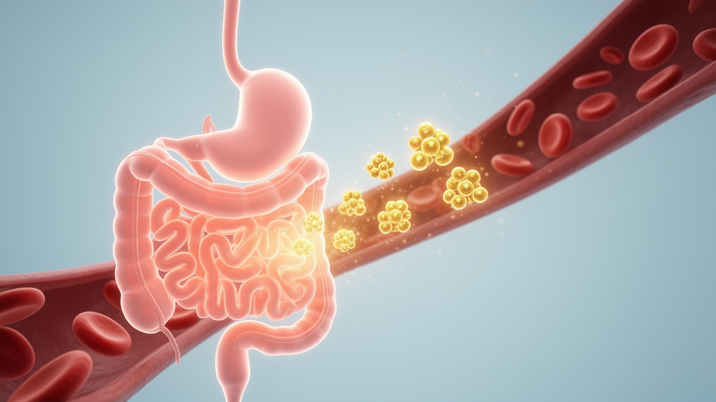
Necrotizing enterocolitis, or NEC, is one of the most feared emergencies in newborn medicine. It strikes roughly 7 to 9 percent of extremely low birth weight infants and is a leading cause of death in babies born before 28 weeks of gestation between postnatal days 15 and 60. The disease begins as injury to the intestinal lining, then can spiral into barrier failure, bacterial translocation, systemic inflammation, and death. Current treatment is largely supportive, and between 20 and 50 percent of affected infants ultimately need surgery because of intestinal perforation or progressive tissue death. Diagnosis still depends on a composite of clinical signs, laboratory tests, and imaging, which means many cases are recognized only after the disease has advanced. A new study published in iScience now reports that a single circulating protein, fibroblast growth factor 5, or FGF5, falls measurably in the blood up to two weeks before NEC is diagnosed, offering a potential route to pre-symptomatic risk stratification in the most fragile preterm infants.
The research team, led by Yu Dai and Shushu Li at Nanjing Women and Children’s Healthcare Hospital, took advantage of an ongoing hospital-based cohort of very preterm infants. Between May 2022 and October 2023, they screened 385 infants born before 32 weeks of gestation and/or weighing under 1,500 grams, of whom 26 developed NEC during hospitalization. Because the cohort stored serum collected on postnatal days 1, 7, 14, and 28, the investigators could retrieve samples drawn within 14 days before each NEC diagnosis. Fifteen NEC infants with such pre-diagnostic samples were matched one-to-one to 15 controls using a twin-sibling priority principle, meaning that when an NEC infant had an unaffected twin, that sibling served as the matched control. After quality control, the final proteomic dataset included 14 NEC cases and 15 controls, with nearly identical gestational ages, birth weights, and perinatal characteristics between groups.
Technically, the study relied on the Olink Target 96 Inflammation Panel, which measures 92 inflammation-related proteins using proximity extension assay technology. In this approach, antibodies against each target carry short DNA tags; when two antibodies bind the same protein in solution, the tags hybridize and are quantified by real-time PCR. The platform outputs normalized protein expression values, or NPX, where one unit corresponds to a twofold difference in protein concentration. This matters for neonatology because blood proteins span several orders of magnitude in abundance and neonatal sample volumes are tiny, constraints that defeat many conventional proteomic platforms. The Olink assay requires only microliters of serum, making serial sampling feasible in infants who weigh little more than a kilogram.
Using paired comparisons between each NEC case and its matched control, the researchers identified four proteins with nominal differences: FGF5, glial cell line-derived neurotrophic factor (GDNF), TRANCE, and uPA. All four were significantly reduced in infants who later developed NEC. After correcting for multiple testing across 92 proteins using the Benjamini-Hochberg false discovery rate, no protein remained significant at q below 0.05, a caveat the authors acknowledge openly. However, when the pre-diagnostic window was split into two strata, 0 to 7 days and 8 to 14 days before diagnosis, FGF5 alone remained persistently downregulated in both intervals, while the other three proteins showed less consistent patterns. Principal component analysis of the full expression matrix showed no clear group separation, which the team interpreted as evidence that NEC-associated differences are subtle and heterogeneous, better captured by matched comparisons than by global variance structure.
The predictive performance of the small protein panel was evaluated with receiver operating characteristic analysis. In the combined 0-to-14-day pre-diagnostic window, FGF5 achieved the highest area under the curve at 0.848, followed by TRANCE at 0.762, GDNF at 0.731, and uPA at 0.710. In the narrower 0-to-7-day window, FGF5’s AUC rose to 0.911, and it maintained 0.781 in the 8-to-14-day window. A Firth-penalized logistic regression model built on pre-diagnostic serum FGF5 reached an AUC of 0.848 with a sensitivity of 0.929 and a specificity of 0.667 at the optimal cutoff. Stratified threefold cross-validation, standard bootstrap resampling with 1,000 iterations, and 0.632+ bootstrap correction all yielded AUC values above 0.840, indicating that the apparent performance was not simply an artifact of model fitting. The authors stress that these analyses remain exploratory given the small cohort and wide confidence intervals.
To strengthen the discovery findings, the team quantified serum FGF5 by enzyme-linked immunosorbent assay in an independent validation cohort of 489 preterm infants prospectively recruited between February 2024 and March 2026. Of the 22 infants diagnosed with NEC in this cohort, 11 had pre-diagnostic serum available and were matched to 11 controls. ELISA verification confirmed significantly lower FGF5 levels before NEC diagnosis, with the direction and magnitude of the between-group difference closely mirroring the Olink data. In this tiny validation set, FGF5 yielded an AUC of 1.0 with perfect sensitivity and specificity, but the authors attribute this to overfitting driven by limited sample size and low between-subject variability, and they explicitly caution against definitive conclusions until larger multicenter cohorts are tested.
What elevates the study beyond a simple biomarker screen is the tissue-level and functional evidence. Because circulating protein levels may not reflect local expression at the site of injury, the investigators examined FGF5 in terminal ileum tissue from surgical resections. Immunohistochemistry and immunofluorescence both showed markedly reduced FGF5 protein in the intestinal mucosa of NEC infants compared with controls, indicating that FGF5 dysregulation occurs at the primary site of pathology and not merely in the circulation. The concordant reduction in serum and tissue supports FGF5 as a biologically relevant marker linked to NEC-associated intestinal injury rather than an incidental correlate of systemic inflammation.
Functional experiments added preliminary mechanistic support. Using the rat small intestinal crypt epithelial cell line IEC-6, the researchers created an inflammatory injury model with lipopolysaccharide treatment and found that both FGF5 protein and mRNA were significantly lower in the NEC-like condition, consistent with the in vivo findings. When they treated the injured cells with 50 nanograms per milliliter of recombinant FGF5 protein, expression of the inflammatory cytokine TNF-alpha fell significantly, suggesting that FGF5 may exert intestinal protective effects by dampening inflammatory signaling. Enrichment analyses of the four candidate proteins pointed in the same direction: Gene Ontology terms clustered around signaling receptor activity, extracellular space localization, cell migration, and cell proliferation, while Kyoto Encyclopedia of Genes and Genomes pathway analysis highlighted the NF-kappaB signaling pathway, calcium signaling, and abnormal cell proliferation as enriched features of NEC progression.
A protein-protein interaction network built from the STRING database placed FGF5 at the center of the candidate signature. Among the 92 measured proteins, which formed 2,535 interaction pairs, FGF5 exhibited the highest connectivity within the candidate subnetwork, with strong predicted associations to GDNF and moderate links to TRANCE and uPA. The authors propose that FGF5 may act as a bifunctional hub, promoting epithelial repair through GDNF-mediated regenerative signaling while modulating inflammatory responses through interactions with TRANCE and uPA. This network position is consistent with the broader biology of the FGF4 subfamily, whose members are implicated in cell proliferation, migration, angiogenesis, and wound repair across multiple organs, although the authors note that NEC-specific mechanisms for FGF5 remain untested and that pathways such as Wnt/beta-catenin and NF-kappaB in FGF5-related NEC biology are literature-based speculation rather than direct findings.
The limitations are substantial and the authors confront them directly. The discovery cohort included only 14 analyzable NEC cases, which diminishes statistical power for screening 92 analytes and elevates the risk of type II errors under multiple testing. As a single-center study, the predictive models retain a high risk of overfitting, an artifact also visible in the small external validation set. Olink proteomics outputs only relative abundance, so standardized clinical cutoffs cannot be derived without dedicated assay development. The IEC-6 cell model, moreover, simplifies NEC-related injury and fails to capture the complexity of prematurity, gut microbiota, perfusion abnormalities, and immune-epithelial interactions that drive the disease in real infants. Still, the pre-diagnostic serial sampling design remains clinically valuable, given the practical barriers to serial blood collection in fragile preterm neonates. If future multicenter studies confirm the signal, a simple blood test measuring FGF5 could one day identify which infants in the neonatal intensive care unit are silently heading toward NEC, opening a window for intervention before the intestine begins to die.
Subject of Research: Necrotizing enterocolitis biomarkers
Article Title: Serum Olink proteomics-based exploration of novel predictive biomarkers for necrotizing enterocolitis
Article References: Dai, Y., Chen, X., Zhang, M., Chen, Y., Yan, X., Chen, X., Li, S., & Han, S. (2026). Serum Olink proteomics-based exploration of novel predictive biomarkers for necrotizing enterocolitis. iScience, 29(11), Article 117617. https://doi.org/10.1016/j.isci.2026.117617
Image Credits: AI Generated
DOI: 10.1016/j.isci.2026.117617
Keywords: proteomics, FGF5, necrotizing enterocolitis, preterm infants, Olink, biomarkers, Serum, proteomics-based, exploration, novel, predictive, necrotizing
Cite Scienmag News
Harold Sullivan. (October 17, 2026). Blood protein FGF5 emerges as early warning sign of deadly infant gut disease. Scienmag. https://scienmag.com/blood-protein-fgf5-emerges-as-early-warning-sign-of-deadly-infant-gut-disease/
Harold Sullivan. "Blood protein FGF5 emerges as early warning sign of deadly infant gut disease." Scienmag, 17 October 2026, https://scienmag.com/blood-protein-fgf5-emerges-as-early-warning-sign-of-deadly-infant-gut-disease/. Accessed 17 October 2026.
Harold Sullivan. "Blood protein FGF5 emerges as early warning sign of deadly infant gut disease." Scienmag. October 17, 2026. https://scienmag.com/blood-protein-fgf5-emerges-as-early-warning-sign-of-deadly-infant-gut-disease/


