


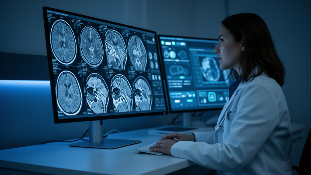
Neuroradiology is undergoing a quiet but profound transformation, one that is reshaping how doctors peer into the living brain and how they decide what to do next. A new collection of studies published in Frontiers in Radiology captures this momentum, spanning artificial intelligence-enhanced CT reconstruction, quantitative MR spectroscopy, vascular imaging biomarkers, and rare pediatric and oncological cases. Together, the seven contributions illustrate a field moving away from purely qualitative pattern recognition and toward objective, threshold-based imaging biomarkers that can be measured, reproduced, and eventually embedded into routine clinical workflows.
The drive to make neuroimaging more quantitative and reproducible runs through much of the collection. One study compared deep learning image reconstruction, or DLIR, with conventional iterative reconstruction in non-contrast head CT. The deep learning approach simultaneously improved gray-white matter differentiation, signal-to-noise and contrast-to-noise ratios, and lesion conspicuity, while allowing a substantial reduction in radiation dose. That last point matters enormously for patients who undergo repeated CT surveillance, where cumulative radiation exposure is a genuine clinical concern. The findings point toward dose-sparing protocols that do not sacrifice diagnostic quality, a trade-off that has long constrained head CT imaging.
Complementing that technical advance, a second study moved quantitative spectroscopy into the diagnostic algorithm for Canavan disease, a rare hereditary white matter disorder. The researchers established cutoff values for NAA/Cr and NAA/Cho ratios on proton MR spectroscopy that achieved excellent diagnostic accuracy in a single-center cohort. Rather than relying on a radiologist’s subjective impression of abnormal spectra, the approach converts metabolic imaging into an objective, threshold-based biomarker. Both studies exemplify the same broader shift: from qualitative pattern recognition toward numbers that can be validated, standardized, and compared across institutions.
Cerebral microbleeds emerged as a unifying imaging marker in two contributions addressing very different vascular substrates. In one case report, a patient with CADASIL, a hereditary cerebral small vessel disease, showed persistent vessel wall enhancement on high-resolution vessel wall MRI that accompanied progression of microbleeds. The observation supports a role for blood-brain barrier disruption in the disease’s pathophysiology and raises the hypothesis that vessel wall imaging could serve as a longitudinal biomarker of disease activity, alongside established markers such as white matter hyperintensity burden. The finding derives from a single case and requires confirmation in larger series, but it hints at a new way to track activity in a disease that currently lacks such tools.
A second contribution took a different angle on microbleeds, synthesizing the heterogeneous literature on their use as markers of traumatic axonal injury after traumatic brain injury. Detected using susceptibility-sensitive sequences such as gradient-echo and susceptibility-weighted imaging, microbleeds have long been proposed as indicators of injury severity, an entity independently characterized using diffusion imaging techniques such as diffusion tensor imaging. The synthesis highlights both the promise and the current inconsistencies in this approach. Read together, the two papers underscore that microbleeds are not a single entity but a context-dependent signal whose interpretation must be anchored to the underlying vascular pathology.
Two contributions addressed the particular challenges of pediatric neuroimaging. One provided a practical guide for the non-pediatric neuroradiologist encountering neonates treated with therapeutic hypothermia for hypoxic-ischemic encephalopathy, summarizing optimal imaging timing, protocols, and characteristic findings. The guidance is particularly valuable as therapeutic hypothermia becomes standard of care in general neonatal units that often lack on-site pediatric neuroradiology expertise. The paper effectively bridges a widening gap between where cooled babies are treated and where specialized imaging interpretation is available.
The second pediatric contribution described a rare presentation of Sturge-Weber syndrome type III in a 5-year-old child without the cutaneous or ocular stigmata typically associated with the syndrome. Multimodal imaging proved decisive for diagnosis despite nonspecific neurological symptoms: CT revealed calcifications, while MRI with susceptibility-weighted and contrast-enhanced sequences identified the leptomeningeal vascular malformation. Both pediatric papers reinforce imaging’s central role when clinical presentation alone is insufficient to guide diagnosis in children, a situation that arises more often than many clinicians appreciate.
Finally, a diagnostically challenging case of gliosarcoma, a rare and aggressive IDH-wildtype glioblastoma variant, demonstrated how subtle imaging features can carry real clinical weight. T2-weighted heterogeneity of the solid tumor component, reflecting the biphasic glial and mesenchymal composition of the tumor, offered a potential radiological clue to a diagnosis that otherwise closely mimics glioblastoma, solitary metastasis, or anaplastic meningioma on conventional sequences. Because gliosarcoma carries a differing prognosis and management pathway relative to its radiological mimics, such imaging features may usefully inform preoperative counselling and surgical planning, even if the observation derives from a single case and needs validation in larger cohorts.
Taken together, the articles reflect the breadth of contemporary neuroradiology, from dose-optimized, AI-assisted CT reconstruction and quantitative spectroscopy to vascular imaging biomarkers spanning hereditary small vessel disease and trauma, and on to the diagnostic value of multimodal imaging in rare pediatric and oncologic presentations. Several directions for future work emerge. Quantitative and AI-based tools will require multicenter, prospective validation before routine incorporation into diagnostic algorithms, together with harmonization of acquisition protocols across scanners and institutions. Imaging biomarkers such as cerebral microbleeds call for pathology-specific interpretive frameworks rather than a one-size-fits-all approach, and would benefit from standardized definitions and reporting criteria to enable comparison across cohorts and diseases.
Rare and atypical presentations, whether a pediatric neurocutaneous syndrome without cutaneous findings or an uncommon glioblastoma variant, continue to depend on the neuroradiologist’s pattern recognition and multimodal integration, even as automated tools mature. Case-based literature remains essential for building the collective experience needed to recognize these entities in daily practice. Looking ahead, translating these findings into clinical benefit will depend on closer collaboration between neuroradiologists, clinicians, and data scientists to embed validated quantitative biomarkers into routine reporting workflows, ultimately supporting earlier diagnosis and more personalized therapeutic strategies for patients with neurological disorders.
Subject of Research: Neuroradiology, artificial intelligence in medical imaging, quantitative imaging biomarkers, and rare disease diagnostics
Article Title: Editorial: Advances in neuroradiology: emerging diagnostic and therapeutic innovations
Article References: Ini’, C., Gigli, S., Mahajan, A., Aliotta, L., & David, E. (2026). Editorial: Advances in neuroradiology: emerging diagnostic and therapeutic innovations. Frontiers in Radiology, 6, Article 1984316. https://doi.org/10.3389/fradi.2026.1984316
Image Credits: AI Generated
DOI: 10.3389/fradi.2026.1984316
Keywords: neuroradiology, artificial intelligence, medical imaging, cerebral microbleeds, pediatric neuroimaging, quantitative imaging, Editorial, Advances, emerging, diagnostic, therapeutic, innovations
Cite Scienmag News
Cassandra Pierce. (October 17, 2026). Neuroradiology’s quiet revolution: from AI-boosted CT scans to smarter brain diagnostics. Scienmag. https://scienmag.com/neuroradiologys-quiet-revolution-from-ai-boosted-ct-scans-to-smarter-brain-diagnostics/
Cassandra Pierce. "Neuroradiology’s quiet revolution: from AI-boosted CT scans to smarter brain diagnostics." Scienmag, 17 October 2026, https://scienmag.com/neuroradiologys-quiet-revolution-from-ai-boosted-ct-scans-to-smarter-brain-diagnostics/. Accessed 17 October 2026.
Cassandra Pierce. "Neuroradiology’s quiet revolution: from AI-boosted CT scans to smarter brain diagnostics." Scienmag. October 17, 2026. https://scienmag.com/neuroradiologys-quiet-revolution-from-ai-boosted-ct-scans-to-smarter-brain-diagnostics/


